2Y0J

Triazoloquinazolines as a novel class of phosphodiesterase 10A (PDE10A) inhibitors, part 2, Lead-optimisation.


Experimental Data Snapshot

  • Method: X-RAY DIFFRACTION
  • Resolution: 2.43 Å
  • R-Value Free: 0.284 
  • R-Value Work: 0.221 
  • R-Value Observed: 0.224 

wwPDB Validation   3D Report Full Report


Ligand Structure Quality Assessment 


This is version 1.2 of the entry. See complete history


Literature

Triazoloquinazolines as a Novel Class of Phosphodiesterase 10A (Pde10A) Inhibitors.

Kehler, J.Ritzen, A.Langgard, M.Petersen, S.L.Farah, M.M.Bundgaard, C.Christoffersen, C.T.Nielsen, J.Kilburn, J.P.

(2011) Bioorg Med Chem Lett 21: 3738

  • DOI: https://doi.org/10.1016/j.bmcl.2011.04.067
  • Primary Citation of Related Structures:  
    2Y0J

  • PubMed Abstract: 

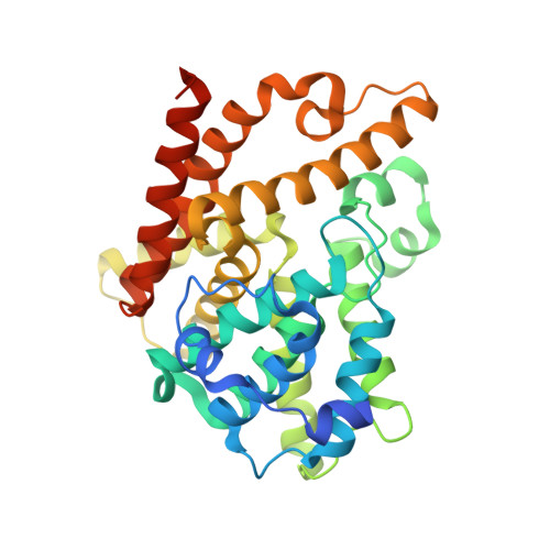
    Novel triazoloquinazolines have been found as phosphodiesterase 10A (PDE10A) inhibitors. Structure-activity studies improved the initial micromolar potency which was found in the lead compound by a 100-fold identifying 5-(1H-benzoimidazol-2-ylmethylsulfanyl)-2-methyl-[1,2,4]triazolo[1,5-c]quinazoline, 42 (PDE10A IC(50)=12 nM) as the most potent compound from the series. Two X-ray structures revealed novel binding modes to the catalytic site of the PDE10A enzyme.


  • Organizational Affiliation

    H. Lundbeck A/S, Department of Discovery Chemistry & DMPK, Valby, Denmark. jke@lundbeck.com


Macromolecules
Find similar proteins by:  (by identity cutoff)  |  3D Structure
Entity ID: 1
MoleculeChains Sequence LengthOrganismDetailsImage
CAMP AND CAMP-INHIBITED CGMP 3', 5'-CYCLIC PHOSPHODIESTERASE 10A
A, B
340Homo sapiensMutation(s): 0 
EC: 3.1.4.17
UniProt & NIH Common Fund Data Resources
Find proteins for Q9Y233 (Homo sapiens)
Explore Q9Y233 
Go to UniProtKB:  Q9Y233
PHAROS:  Q9Y233
GTEx:  ENSG00000112541 
Entity Groups  
Sequence Clusters30% Identity50% Identity70% Identity90% Identity95% Identity100% Identity
UniProt GroupQ9Y233
Sequence Annotations
Expand
  • Reference Sequence
Small Molecules
Binding Affinity Annotations 
IDSourceBinding Affinity
AXC PDBBind:  2Y0J IC50: 12 (nM) from 1 assay(s)
BindingDB:  2Y0J IC50: 12 (nM) from 1 assay(s)
Binding MOAD:  2Y0J IC50: 12 (nM) from 1 assay(s)
Experimental Data & Validation

Experimental Data

  • Method: X-RAY DIFFRACTION
  • Resolution: 2.43 Å
  • R-Value Free: 0.284 
  • R-Value Work: 0.221 
  • R-Value Observed: 0.224 
  • Space Group: P 21 21 21
Unit Cell:
Length ( Å )Angle ( ˚ )
a = 49.38α = 90
b = 81.69β = 90
c = 160.62γ = 90
Software Package:
Software NamePurpose
REFMACrefinement
MOSFLMdata reduction
SCALAdata scaling
MOLREPphasing

Structure Validation

View Full Validation Report


Ligand Structure Quality Assessment 


Entry History 

Deposition Data

Revision History  (Full details and data files)

  • Version 1.0: 2011-06-15
    Type: Initial release
  • Version 1.1: 2011-07-13
    Changes: Version format compliance
  • Version 1.2: 2023-12-20
    Changes: Data collection, Database references, Derived calculations, Other, Refinement description