2XXG

STRUCTURE OF THE N90S MUTANT OF NITRITE REDUCTASE FROM ALCALIGENES XYLOSOXIDANS


Experimental Data Snapshot

  • Method: X-RAY DIFFRACTION
  • Resolution: 1.60 Å
  • R-Value Free: 0.211 
  • R-Value Work: 0.168 
  • R-Value Observed: 0.170 

wwPDB Validation   3D Report Full Report


This is version 2.1 of the entry. See complete history


Literature

Identification of the Proton Channel to the Active Site Type 2 Cu Centre of Nitrite Reductase: Structural and Enzymatic Properties of the His254Phe and Asn90Ser Mutants

Hough, M.A.Eady, R.R.Hasnain, S.S.

(2008) Biochemistry 47: 13547

  • DOI: https://doi.org/10.1021/bi801369y
  • Primary Citation of Related Structures:  
    2XXG

  • PubMed Abstract: 

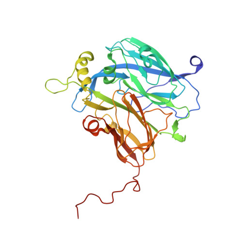
    Proton and electron delivery to the catalytic site and their associated pathways are crucial elements in understanding the mechanisms of redox enzymes. Two distinct proton channels have previously been identified in copper nitrite reductases based on high- to atomic-resolution crystal structures. These were assigned as the "primary" and "high-pH" proton channels and link the catalytic type 2 Cu center to the enzyme surface. Residue His254 has been identified as a key residue in the primary proton channel from the catalytic T2Cu site to the surface, while Asn90 is thought to be a key residue in the high-pH channel. The structure of the His254Phe mutant was previously determined to 1.85 A resolution, revealing disruption in the H-bonding network of the primary proton channel. The effect of the mutation on proton transfer was not established as the T2Cu center was unusually occupied by Zn. New growth protocols have now led to the incorporation of copper at this site, and here we present spectroscopic, catalytic activity, and structural data for the Cu-loaded H254F mutant of AxNiR. Surprisingly, this species exhibits essentially full catalytic activity, despite the clear disruption of the primary proton channel. In contrast, the Asn90Ser mutation disrupts H-bonding in the high-pH proton channel and results in an approximately 70% decrease in specific activity. These mutations do not change the apparent K(m) for nitrite, and thus, these data clearly demonstrate a role for the high-pH proton channel in the delivery of protons to the catalytic T2Cu center at physiological pH values; it may in fact be the main source of protons to the T2Cu center.


  • Organizational Affiliation

    Molecular Biophysics Group, School of Biological Sciences, University of Liverpool, UK.


Macromolecules
Find similar proteins by:  (by identity cutoff)  |  3D Structure
Entity ID: 1
MoleculeChains Sequence LengthOrganismDetailsImage
DISSIMILATORY COPPER-CONTAINING NITRITE REDUCTASEA,
B [auth C]
336Achromobacter xylosoxidansMutation(s): 1 
UniProt
Find proteins for O68601 (Alcaligenes xylosoxydans xylosoxydans)
Explore O68601 
Go to UniProtKB:  O68601
Entity Groups  
Sequence Clusters30% Identity50% Identity70% Identity90% Identity95% Identity100% Identity
UniProt GroupO68601
Sequence Annotations
Expand
  • Reference Sequence
Small Molecules
Ligands 5 Unique
IDChains Name / Formula / InChI Key2D Diagram3D Interactions
MES
Query on MES

Download Ideal Coordinates CCD File 
I [auth A],
J [auth A],
S [auth C],
T [auth C]
2-(N-MORPHOLINO)-ETHANESULFONIC ACID
C6 H13 N O4 S
SXGZJKUKBWWHRA-UHFFFAOYSA-N
PG4
Query on PG4

Download Ideal Coordinates CCD File 
C [auth A],
L [auth C]
TETRAETHYLENE GLYCOL
C8 H18 O5
UWHCKJMYHZGTIT-UHFFFAOYSA-N
PEG
Query on PEG

Download Ideal Coordinates CCD File 
G [auth A],
K [auth A],
M [auth C],
Q [auth C]
DI(HYDROXYETHYL)ETHER
C4 H10 O3
MTHSVFCYNBDYFN-UHFFFAOYSA-N
ZN
Query on ZN

Download Ideal Coordinates CCD File 
F [auth A],
H [auth A],
P [auth C],
R [auth C]
ZINC ION
Zn
PTFCDOFLOPIGGS-UHFFFAOYSA-N
CU
Query on CU

Download Ideal Coordinates CCD File 
D [auth A],
E [auth A],
N [auth C],
O [auth C]
COPPER (II) ION
Cu
JPVYNHNXODAKFH-UHFFFAOYSA-N
Modified Residues  1 Unique
IDChains TypeFormula2D DiagramParent
PCA
Query on PCA
A,
B [auth C]
L-PEPTIDE LINKINGC5 H7 N O3GLN
Experimental Data & Validation

Experimental Data

  • Method: X-RAY DIFFRACTION
  • Resolution: 1.60 Å
  • R-Value Free: 0.211 
  • R-Value Work: 0.168 
  • R-Value Observed: 0.170 
  • Space Group: H 3
Unit Cell:
Length ( Å )Angle ( ˚ )
a = 89.552α = 90
b = 89.552β = 90
c = 287.678γ = 120
Software Package:
Software NamePurpose
REFMACrefinement
HKL-2000data reduction
HKL-2000data scaling
PHASERphasing

Structure Validation

View Full Validation Report


Entry History 

Deposition Data

Revision History  (Full details and data files)

  • Version 1.0: 2011-05-18
    Type: Initial release
  • Version 1.1: 2011-07-13
    Changes: Version format compliance
  • Version 2.0: 2020-03-11
    Changes: Derived calculations, Other, Polymer sequence
  • Version 2.1: 2023-12-20
    Changes: Data collection, Database references, Derived calculations, Refinement description