2XWT

CRYSTAL STRUCTURE OF THE TSH RECEPTOR IN COMPLEX WITH A BLOCKING TYPE TSHR AUTOANTIBODY


Experimental Data Snapshot

  • Method: X-RAY DIFFRACTION
  • Resolution: 1.90 Å
  • R-Value Free: 0.227 
  • R-Value Work: 0.177 
  • R-Value Observed: 0.179 

wwPDB Validation   3D Report Full Report


Ligand Structure Quality Assessment 


This is version 2.1 of the entry. See complete history


Literature

Crystal Structure of the Tsh Receptor (Tshr) Bound to a Blocking-Type Tshr Autoantibody.

Sanders, P.Young, S.Sanders, J.Kabelis, K.Baker, S.Sullivan, A.Evans, M.Clark, J.Wilmot, J.Hu, X.Roberts, E.Powell, M.Nunez Miguel, R.Furmaniak, J.Rees Smith, B.

(2011) J Mol Endocrinol 46: 81

  • DOI: https://doi.org/10.1530/JME-10-0127
  • Primary Citation of Related Structures:  
    2XWT

  • PubMed Abstract: 

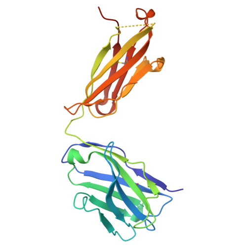
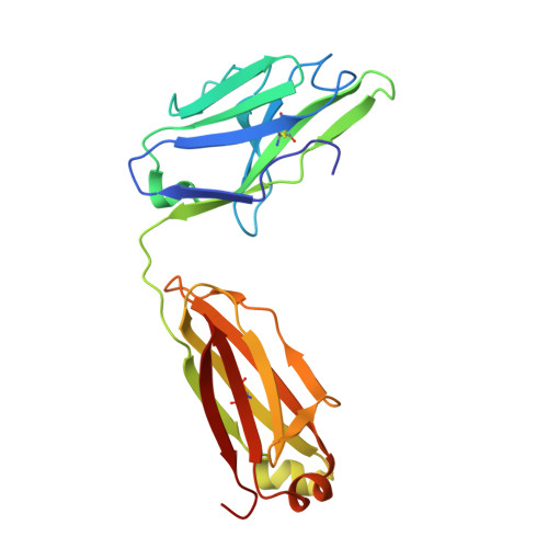
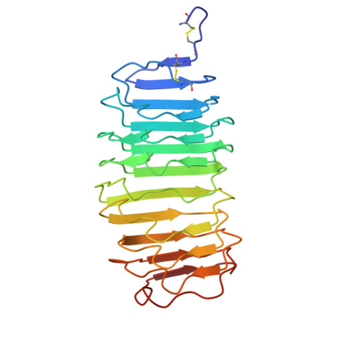
    A complex of the TSH receptor extracellular domain (amino acids 22-260; TSHR260) bound to a blocking-type human monoclonal autoantibody (K1-70) was purified, crystallised and the structure solved at 1.9 Å resolution. K1-70 Fab binds to the concave surface of the TSHR leucine-rich domain (LRD) forming a large interface (2565 Å(2)) with an extensive network of ionic, polar and hydrophobic interactions. Mutation of TSHR or K1-70 residues showing strong interactions in the solved structure influenced the activity of K1-70, indicating that the binding detail observed in the complex reflects interactions of K1-70 with intact, functionally active TSHR. Unbound K1-70 Fab was prepared and crystallised to 2.22 Å resolution. Virtually no movement was observed in the atoms of K1-70 residues on the binding interface compared with unbound K1-70, consistent with 'lock and key' binding. The binding arrangements in the TSHR260-K1-70 Fab complex are similar to previously observed for the TSHR260-M22 Fab complex; however, K1-70 clasps the concave surface of the TSHR LRD in approximately the opposite orientation (rotated 155°) to M22. The blocking autoantibody K1-70 binds more N-terminally on the TSHR concave surface than either the stimulating autoantibody M22 or the hormone TSH, and this may reflect its different functional activity. The structure of TSHR260 in the TSHR260-K1-70 and TSHR260-M22 complexes show a root mean square deviation on all C(α) atoms of only 0.51 Å. These high-resolution crystal structures provide a foundation for developing new strategies to understand and control TSHR activation and the autoimmune response to the TSHR.


  • Organizational Affiliation

    FIRS Laboratories, RSR Ltd, Parc Ty Glas, Llanishen, Cardiff CF14 5DU, UK.


Macromolecules
Find similar proteins by:  (by identity cutoff)  |  3D Structure
Entity ID: 1
MoleculeChains Sequence LengthOrganismDetailsImage
THYROID BLOCKING HUMAN AUTOANTIBODY K1-70 HEAVY CHAIN221Homo sapiensMutation(s): 0 
Entity Groups  
Sequence Clusters30% Identity50% Identity70% Identity90% Identity95% Identity100% Identity
Sequence Annotations
Expand
  • Reference Sequence
Find similar proteins by:  (by identity cutoff)  |  3D Structure
Entity ID: 2
MoleculeChains Sequence LengthOrganismDetailsImage
THYROID BLOCKING HUMAN AUTOANTIBODY K1-70 LIGHT CHAIN214Homo sapiensMutation(s): 0 
Entity Groups  
Sequence Clusters30% Identity50% Identity70% Identity90% Identity95% Identity100% Identity
Sequence Annotations
Expand
  • Reference Sequence
Find similar proteins by:  (by identity cutoff)  |  3D Structure
Entity ID: 3
MoleculeChains Sequence LengthOrganismDetailsImage
THYROTROPIN RECEPTOR239Homo sapiensMutation(s): 0 
UniProt & NIH Common Fund Data Resources
Find proteins for P16473 (Homo sapiens)
Explore P16473 
Go to UniProtKB:  P16473
PHAROS:  P16473
GTEx:  ENSG00000165409 
Entity Groups  
Sequence Clusters30% Identity50% Identity70% Identity90% Identity95% Identity100% Identity
UniProt GroupP16473
Sequence Annotations
Expand
  • Reference Sequence
Oligosaccharides

Help

Entity ID: 4
MoleculeChains Length2D Diagram Glycosylation3D Interactions
2-acetamido-2-deoxy-beta-D-glucopyranose-(1-4)-2-acetamido-2-deoxy-beta-D-glucopyranose
D
2N-Glycosylation
Glycosylation Resources
GlyTouCan:  G42666HT
GlyCosmos:  G42666HT
GlyGen:  G42666HT
Entity ID: 5
MoleculeChains Length2D Diagram Glycosylation3D Interactions
alpha-D-mannopyranose-(1-6)-beta-D-mannopyranose-(1-4)-2-acetamido-2-deoxy-beta-D-glucopyranose-(1-4)-2-acetamido-2-deoxy-beta-D-glucopyranose
E
4N-Glycosylation
Glycosylation Resources
GlyTouCan:  G22573RC
GlyCosmos:  G22573RC
GlyGen:  G22573RC
Small Molecules
Ligands 1 Unique
IDChains Name / Formula / InChI Key2D Diagram3D Interactions
NAG
Query on NAG

Download Ideal Coordinates CCD File 
F [auth C],
G [auth C]
2-acetamido-2-deoxy-beta-D-glucopyranose
C8 H15 N O6
OVRNDRQMDRJTHS-FMDGEEDCSA-N
Experimental Data & Validation

Experimental Data

  • Method: X-RAY DIFFRACTION
  • Resolution: 1.90 Å
  • R-Value Free: 0.227 
  • R-Value Work: 0.177 
  • R-Value Observed: 0.179 
  • Space Group: P 21 21 21
Unit Cell:
Length ( Å )Angle ( ˚ )
a = 83.02α = 90
b = 89.3β = 90
c = 101.27γ = 90
Software Package:
Software NamePurpose
REFMACrefinement
MOSFLMdata reduction
SCALAdata scaling
PHASERphasing

Structure Validation

View Full Validation Report


Ligand Structure Quality Assessment 


Entry History 

Revision History  (Full details and data files)

  • Version 1.0: 2011-03-09
    Type: Initial release
  • Version 1.1: 2011-05-08
    Changes: Version format compliance
  • Version 1.2: 2011-07-13
    Changes: Version format compliance
  • Version 1.3: 2017-06-28
    Changes: Data collection
  • Version 1.4: 2019-04-03
    Changes: Data collection, Source and taxonomy
  • Version 2.0: 2020-07-29
    Type: Remediation
    Reason: Carbohydrate remediation
    Changes: Atomic model, Data collection, Derived calculations, Other, Structure summary
  • Version 2.1: 2023-12-20
    Changes: Data collection, Database references, Derived calculations, Refinement description, Structure summary