2XVA

Crystal structure of the tellurite detoxification protein TehB from E. coli in complex with sinefungin


Experimental Data Snapshot

  • Method: X-RAY DIFFRACTION
  • Resolution: 1.90 Å
  • R-Value Free: 0.234 
  • R-Value Work: 0.175 
  • R-Value Observed: 0.178 

wwPDB Validation   3D Report Full Report


Ligand Structure Quality Assessment 


This is version 1.2 of the entry. See complete history


Literature

Structure and Mechanism of the Chalcogen Detoxifying Protein Tehb from Escherichia Coli.

Choudhury, H.G.Cameron, A.D.Iwata, S.Beis, K.

(2011) Biochem J 435: 85

  • DOI: https://doi.org/10.1042/BJ20102014
  • Primary Citation of Related Structures:  
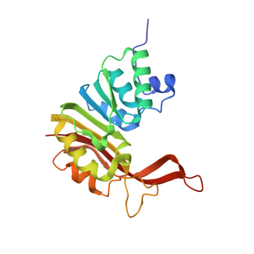
    2XVA, 2XVM

  • PubMed Abstract: 

    The oxyanion derivatives of the chalcogens tellurium and selenium are toxic to living organisms even at very low levels. Bacteria have developed mechanisms to overcome their toxicity by methylating them. The structure of TehB from Escherichia coli has been determined in the presence of the cofactor analogues SAH (S-adenosylhomocysteine) and sinefungin (a non-hydrolysable form of S-adenosyl-L-methionine) at 1.48 Å (1 Å=0.1 nm) and 1.9 Å respectively. Interestingly, our kinetic data show that TehB does not discriminate between selenium or tellurite oxyanions, making it a very powerful detoxifying protein. Analysis of the active site has identified three conserved residues that are capable of binding and orientating the metals for nucleophilic attack: His176, Arg177 and Arg184. Mutagenesis studies revealed that the H176A and R184A mutants retained most of their activity, whereas the R177A mutant had 65% of its activity abolished. Based on the structure and kinetic data we propose an SN2 nucleophilic attack reaction mechanism. These data provide the first molecular understanding of the detoxification of chalcogens by bacteria.


  • Organizational Affiliation

    Membrane Protein Laboratory, Imperial College London, Diamond Light Source, Chilton, Oxfordshire OX11 0DE, UK.


Macromolecules
Find similar proteins by:  (by identity cutoff)  |  3D Structure
Entity ID: 1
MoleculeChains Sequence LengthOrganismDetailsImage
TELLURITE RESISTANCE PROTEIN TEHB
A, B, C, D
199Escherichia coli str. K-12 substr. MG1655Mutation(s): 0 
UniProt
Find proteins for P25397 (Escherichia coli (strain K12))
Explore P25397 
Go to UniProtKB:  P25397
Entity Groups  
Sequence Clusters30% Identity50% Identity70% Identity90% Identity95% Identity100% Identity
UniProt GroupP25397
Sequence Annotations
Expand
  • Reference Sequence
Experimental Data & Validation

Experimental Data

  • Method: X-RAY DIFFRACTION
  • Resolution: 1.90 Å
  • R-Value Free: 0.234 
  • R-Value Work: 0.175 
  • R-Value Observed: 0.178 
  • Space Group: P 1 21 1
Unit Cell:
Length ( Å )Angle ( ˚ )
a = 59.1α = 90
b = 55.59β = 95.93
c = 129.7γ = 90
Software Package:
Software NamePurpose
REFMACrefinement
XDSdata reduction
SCALAdata scaling
PHASERphasing

Structure Validation

View Full Validation Report


Ligand Structure Quality Assessment 


Entry History 

Deposition Data

Revision History  (Full details and data files)

  • Version 1.0: 2011-02-16
    Type: Initial release
  • Version 1.1: 2012-09-05
    Changes: Derived calculations, Non-polymer description, Other, Structure summary, Version format compliance
  • Version 1.2: 2023-12-20
    Changes: Data collection, Database references, Derived calculations, Other, Refinement description