2XQ1

Crystal structure of peroxisomal catalase from the yeast Hansenula polymorpha


Experimental Data Snapshot

  • Method: X-RAY DIFFRACTION
  • Resolution: 2.90 Å
  • R-Value Free: 0.223 
  • R-Value Work: 0.191 
  • R-Value Observed: 0.191 

wwPDB Validation   3D Report Full Report


Ligand Structure Quality Assessment 


This is version 1.3 of the entry. See complete history


Literature

Structural Features of Peroxisomal Catalase from the Yeast Hansenula Polymorpha

Penya-Soler, E.Vega, M.C.Wilmanns, M.Williams, C.P.

(2011) Acta Crystallogr D Biol Crystallogr 67: 690

  • DOI: https://doi.org/10.1107/S0907444911022463
  • Primary Citation of Related Structures:  
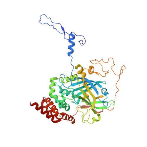
    2XQ1

  • PubMed Abstract: 

    The reactive oxygen species hydrogen peroxide is a byproduct of the β-oxidation process that occurs in peroxisomes. Since reactive oxygen species can cause serious damage to biomolecules, a number of scavengers control their intracellular levels. One such scavenger that is present in the peroxisome is the oxidoreductase catalase. In this study, the crystal structure of heterologously expressed peroxisomal catalase from the thermotolerant yeast Hansenula polymorpha has been determined at 2.9 Å resolution. H. polymorpha catalase is a typical peroxisomal catalase; it is tetrameric and is highly similar to catalases from other organisms. However, its hydrogen peroxide-degrading activity is higher than those of a number of other catalases for which structural data are available. Structural superimpositions indicate that the nature of the major channel, the path for hydrogen peroxide to the active site, varies from those seen in other catalase structures, an observation that may account for the high activity of H. polymorpha catalase.


  • Organizational Affiliation

    Department of Structural and Quantitative Biology, Centro de Investigaciones Biológicas (CIB-CSIC), Ramiro de Maetzu, 28040 Madrid, Spain.


Macromolecules
Find similar proteins by:  (by identity cutoff)  |  3D Structure
Entity ID: 1
MoleculeChains Sequence LengthOrganismDetailsImage
PEROXISOMAL CATALASE
A, B, C, D, E
A, B, C, D, E, F, G, H, I, J, K, L, M, N, O, P
509Ogataea angustaMutation(s): 0 
EC: 1.11.1.6
UniProt
Find proteins for P30263 (Pichia angusta)
Explore P30263 
Go to UniProtKB:  P30263
Entity Groups  
Sequence Clusters30% Identity50% Identity70% Identity90% Identity95% Identity100% Identity
UniProt GroupP30263
Sequence Annotations
Expand
  • Reference Sequence
Small Molecules
Ligands 1 Unique
IDChains Name / Formula / InChI Key2D Diagram3D Interactions
HEM
Query on HEM

Download Ideal Coordinates CCD File 
AA [auth L]
BA [auth M]
CA [auth N]
DA [auth O]
EA [auth P]
AA [auth L],
BA [auth M],
CA [auth N],
DA [auth O],
EA [auth P],
Q [auth B],
R [auth C],
S [auth D],
T [auth E],
U [auth F],
V [auth G],
W [auth H],
X [auth I],
Y [auth J],
Z [auth K]
PROTOPORPHYRIN IX CONTAINING FE
C34 H32 Fe N4 O4
KABFMIBPWCXCRK-RGGAHWMASA-L
Experimental Data & Validation

Experimental Data

  • Method: X-RAY DIFFRACTION
  • Resolution: 2.90 Å
  • R-Value Free: 0.223 
  • R-Value Work: 0.191 
  • R-Value Observed: 0.191 
  • Space Group: P 1 21 1
Unit Cell:
Length ( Å )Angle ( ˚ )
a = 132.52α = 90
b = 196.68β = 92.85
c = 170.85γ = 90
Software Package:
Software NamePurpose
REFMACrefinement
XDSdata reduction
SCALAdata scaling
MOLREPphasing

Structure Validation

View Full Validation Report


Ligand Structure Quality Assessment 


Entry History 

Deposition Data

Revision History  (Full details and data files)

  • Version 1.0: 2011-06-22
    Type: Initial release
  • Version 1.1: 2011-07-27
    Changes: Database references, Version format compliance
  • Version 1.2: 2012-03-28
    Changes: Other
  • Version 1.3: 2023-12-20
    Changes: Data collection, Database references, Derived calculations, Other, Refinement description