2XE4

Structure of Oligopeptidase B from Leishmania major


Experimental Data Snapshot

  • Method: X-RAY DIFFRACTION
  • Resolution: 1.65 Å
  • R-Value Free: 0.178 
  • R-Value Work: 0.140 
  • R-Value Observed: 0.142 

wwPDB Validation   3D Report Full Report


This is version 2.0 of the entry. See complete history


Literature

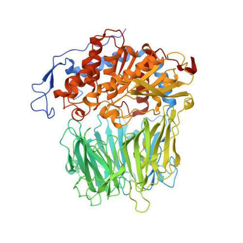
Crystal Structure of Leishmania Major Oligopeptidase B Gives Insight Into the Enzymatic Properties of a Trypanosomatid Virulence Factor.

Mcluskey, K.Paterson, N.G.Bland, N.D.Isaacs, N.W.Mottram, J.C.

(2010) J Biol Chem 285: 39249

  • DOI: https://doi.org/10.1074/jbc.M110.156679
  • Primary Citation of Related Structures:  
    2XE4

  • PubMed Abstract: 

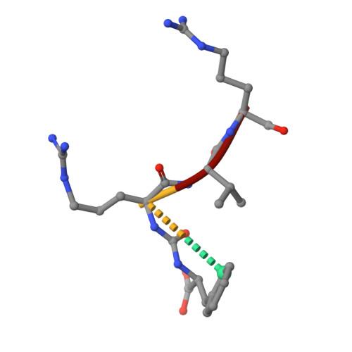
    Oligopeptidase B (OPB) is a serine peptidase with dibasic substrate specificity. It is found in bacteria, plants, and trypanosomatid pathogens, where it has been identified as a virulence factor and potential drug target. In this study we expressed active recombinant Leishmania major OPB and provide the first structure of an oligopeptidase B at high resolution. The crystallographic study reveals that OPB comprises two domains, a catalytic and a propeller domain, linked together by a hinge region. The structure has been determined in complex with the oligopeptide, protease-inhibitor antipain, giving detailed information on the enzyme active site and extended substrate binding pockets. It shows that Glu-621 plays a critical role in the S1 binding pocket and, along with Phe-603, is largely responsible for the enzyme substrate specificity in P1. In the S2 binding pocket, Tyr-499 was shown to be important for substrate stability. The structure also allowed an investigation into the function of residues highlighted in other studies including Glu-623, which was predicted to be involved in the S1 binding pocket but is found forming an inter-domain hydrogen bond. Additional important salt bridges/hydrogen bonds between the two domains were observed, highlighting the significance of the domain interface in OPB. This work provides a foundation for the study of the role of OPBs as virulence factors in trypanosomatids. It could facilitate the development of specific OPB inhibitors with therapeutic potential by exploiting its unique substrate recognition properties as well as providing a model for OPBs in general.


  • Organizational Affiliation

    Westchem School of Chemistry, University of Glasgow, Glasgow G12 8TA, Scotland, United Kingdom. Karen.McLuskey@glasgow.ac.uk


Macromolecules
Find similar proteins by:  (by identity cutoff)  |  3D Structure
Entity ID: 1
MoleculeChains Sequence LengthOrganismDetailsImage
OLIGOPEPTIDASE B751Leishmania majorMutation(s): 1 
EC: 3.4.21.83
UniProt
Find proteins for Q4QHU7 (Leishmania major)
Explore Q4QHU7 
Go to UniProtKB:  Q4QHU7
Entity Groups  
Sequence Clusters30% Identity50% Identity70% Identity90% Identity95% Identity100% Identity
UniProt GroupQ4QHU7
Sequence Annotations
Expand
  • Reference Sequence

Find similar proteins by:  Sequence   |   3D Structure  

Entity ID: 2
MoleculeChains Sequence LengthOrganismDetailsImage
ANTIPAIN4ActinomycetesMutation(s): 0 
Sequence Annotations
Expand
  • Reference Sequence
Small Molecules
Ligands 6 Unique
IDChains Name / Formula / InChI Key2D Diagram3D Interactions
PO4
Query on PO4

Download Ideal Coordinates CCD File 
JB [auth A],
KB [auth A],
LB [auth A]
PHOSPHATE ION
O4 P
NBIIXXVUZAFLBC-UHFFFAOYSA-K
GOL
Query on GOL

Download Ideal Coordinates CCD File 
C [auth A]
D [auth A]
E [auth A]
F [auth A]
G [auth A]
C [auth A],
D [auth A],
E [auth A],
F [auth A],
G [auth A],
H [auth A],
I [auth A],
J [auth A],
K [auth A],
L [auth A]
GLYCEROL
C3 H8 O3
PEDCQBHIVMGVHV-UHFFFAOYSA-N
PGO
Query on PGO

Download Ideal Coordinates CCD File 
N [auth A]S-1,2-PROPANEDIOL
C3 H8 O2
DNIAPMSPPWPWGF-VKHMYHEASA-N
PGR
Query on PGR

Download Ideal Coordinates CCD File 
AA [auth A]
AB [auth A]
BA [auth A]
BB [auth A]
CA [auth A]
AA [auth A],
AB [auth A],
BA [auth A],
BB [auth A],
CA [auth A],
CB [auth A],
DA [auth A],
DB [auth A],
EA [auth A],
EB [auth A],
FA [auth A],
FB [auth A],
GA [auth A],
GB [auth A],
HA [auth A],
HB [auth A],
IA [auth A],
IB [auth A],
JA [auth A],
KA [auth A],
LA [auth A],
M [auth A],
MA [auth A],
NA [auth A],
O [auth A],
OA [auth A],
P [auth A],
PA [auth A],
Q [auth A],
QA [auth A],
R [auth A],
RA [auth A],
S [auth A],
SA [auth A],
T [auth A],
TA [auth A],
U [auth A],
UA [auth A],
V [auth A],
VA [auth A],
W [auth A],
WA [auth A],
X [auth A],
XA [auth A],
Y [auth A],
YA [auth A],
Z [auth A],
ZA [auth A]
R-1,2-PROPANEDIOL
C3 H8 O2
DNIAPMSPPWPWGF-GSVOUGTGSA-N
CL
Query on CL

Download Ideal Coordinates CCD File 
MB [auth A],
OB [auth A],
QB [auth A],
RB [auth A]
CHLORIDE ION
Cl
VEXZGXHMUGYJMC-UHFFFAOYSA-M
NA
Query on NA

Download Ideal Coordinates CCD File 
NB [auth A],
PB [auth A]
SODIUM ION
Na
FKNQFGJONOIPTF-UHFFFAOYSA-N
Modified Residues  1 Unique
IDChains TypeFormula2D DiagramParent
RGL
Query on RGL
B
L-PEPTIDE LINKINGC6 H15 N4 OARG
Biologically Interesting Molecules (External Reference) 1 Unique
Experimental Data & Validation

Experimental Data

  • Method: X-RAY DIFFRACTION
  • Resolution: 1.65 Å
  • R-Value Free: 0.178 
  • R-Value Work: 0.140 
  • R-Value Observed: 0.142 
  • Space Group: I 2 2 2
Unit Cell:
Length ( Å )Angle ( ˚ )
a = 95.478α = 90
b = 142.776β = 90
c = 208.919γ = 90
Software Package:
Software NamePurpose
REFMACrefinement
d*TREKdata reduction
SCALAdata scaling
autoSHARPphasing
SHELXCDphasing
SHELXDphasing

Structure Validation

View Full Validation Report


Entry History 

Deposition Data

Revision History  (Full details and data files)

  • Version 1.0: 2010-10-06
    Type: Initial release
  • Version 1.1: 2011-08-10
    Changes: Database references, Structure summary, Version format compliance
  • Version 1.2: 2012-11-30
    Changes: Other
  • Version 1.3: 2013-08-07
    Changes: Other
  • Version 2.0: 2019-05-15
    Changes: Advisory, Data collection, Derived calculations, Other, Polymer sequence