2X56

Yersinia Pestis Plasminogen Activator Pla (Native)


Experimental Data Snapshot

  • Method: X-RAY DIFFRACTION
  • Resolution: 2.30 Å
  • R-Value Free: 0.226 
  • R-Value Work: 0.199 
  • R-Value Observed: 0.201 

wwPDB Validation   3D Report Full Report


Ligand Structure Quality Assessment 


This is version 1.2 of the entry. See complete history


Literature

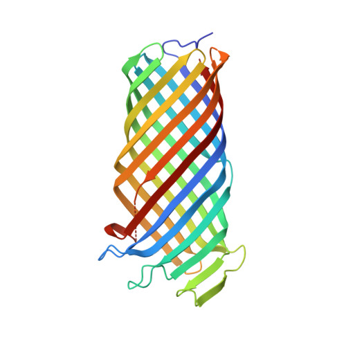
An Active Site Water Network in the Plasminogen Activator Pla from Yersinia Pestis

Eren, E.Murphy, M.Goguen, J.van den Berg, B.

(2010) Structure 18: 809

  • DOI: https://doi.org/10.1016/j.str.2010.03.013
  • Primary Citation of Related Structures:  
    2X4M, 2X55, 2X56

  • PubMed Abstract: 

    The plasminogen activator Pla from Yersinia pestis is an outer membrane protease (omptin) that is important for the virulence of plague. Here, we present the high-resolution crystal structure of wild-type, enzymatically active Pla at 1.9 A. The structure shows a water molecule located between active site residues D84 and H208, which likely corresponds to the nucleophilic water. A number of other water molecules are present in the active site, linking residues important for enzymatic activity. The R211 sidechain in loop L4 is close to the nucleophilic water and possibly involved in the stabilization of the oxyanion intermediate. Subtle conformational changes of H208 result from the binding of lipopolysaccharide to the outside of the barrel, explaining the unusual dependence of omptins on lipopolysaccharide for activity. The Pla structure suggests a model for the interaction with plasminogen substrate and provides a more detailed understanding of the catalytic mechanism of omptin proteases.


  • Organizational Affiliation

    Program in Molecular Medicine, University of Massachusetts Medical School, Worcester, MA 01605, USA.


Macromolecules
Find similar proteins by:  (by identity cutoff)  |  3D Structure
Entity ID: 1
MoleculeChains Sequence LengthOrganismDetailsImage
COAGULASE/FIBRINOLYSIN292Yersinia pestisMutation(s): 0 
EC: 3.4.23.48
Membrane Entity: Yes 
UniProt
Find proteins for P17811 (Yersinia pestis)
Explore P17811 
Go to UniProtKB:  P17811
Entity Groups  
Sequence Clusters30% Identity50% Identity70% Identity90% Identity95% Identity100% Identity
UniProt GroupP17811
Sequence Annotations
Expand
  • Reference Sequence
Small Molecules
Ligands 1 Unique
IDChains Name / Formula / InChI Key2D Diagram3D Interactions
C8E
Query on C8E

Download Ideal Coordinates CCD File 
B [auth A]
C [auth A]
D [auth A]
E [auth A]
F [auth A]
B [auth A],
C [auth A],
D [auth A],
E [auth A],
F [auth A],
G [auth A],
H [auth A],
I [auth A],
J [auth A],
K [auth A],
L [auth A],
M [auth A],
N [auth A],
O [auth A],
P [auth A],
Q [auth A],
R [auth A],
S [auth A]
(HYDROXYETHYLOXY)TRI(ETHYLOXY)OCTANE
C16 H34 O5
FEOZZFHAVXYAMB-UHFFFAOYSA-N
Experimental Data & Validation

Experimental Data

  • Method: X-RAY DIFFRACTION
  • Resolution: 2.30 Å
  • R-Value Free: 0.226 
  • R-Value Work: 0.199 
  • R-Value Observed: 0.201 
  • Space Group: P 65 2 2
Unit Cell:
Length ( Å )Angle ( ˚ )
a = 140.241α = 90
b = 140.241β = 90
c = 130.502γ = 120
Software Package:
Software NamePurpose
PHENIXrefinement
HKL-2000data reduction
SCALEPACKdata scaling
PHASERphasing

Structure Validation

View Full Validation Report


Ligand Structure Quality Assessment 


Entry History 

Deposition Data

Revision History  (Full details and data files)

  • Version 1.0: 2010-07-28
    Type: Initial release
  • Version 1.1: 2012-03-28
    Changes: Database references, Derived calculations, Refinement description, Version format compliance
  • Version 1.2: 2018-01-24
    Changes: Database references, Source and taxonomy