2WRH

structure of H1 duck albert hemagglutinin with human receptor


Experimental Data Snapshot

  • Method: X-RAY DIFFRACTION
  • Resolution: 3.00 Å
  • R-Value Free: 0.252 
  • R-Value Work: 0.229 
  • R-Value Observed: 0.230 

wwPDB Validation   3D Report Full Report


Ligand Structure Quality Assessment 


This is version 1.2 of the entry. See complete history


Literature

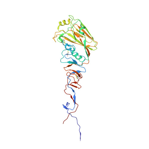
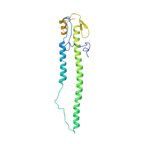
Structures of Receptor Complexes Formed by Hemagglutinins from the Asian Influenza Pandemic of 1957

Liu, J.Stevens, D.J.Haire, L.F.Walker, P.A.Coombs, P.J.Russell, R.J.Gamblin, S.J.Skehel, J.J.

(2009) Proc Natl Acad Sci U S A 106: 17175

  • DOI: https://doi.org/10.1073/pnas.0906849106
  • Primary Citation of Related Structures:  
    2WR0, 2WR1, 2WR2, 2WR3, 2WR4, 2WR5, 2WR7, 2WRB, 2WRC, 2WRD, 2WRE, 2WRF, 2WRG, 2WRH

  • PubMed Abstract: 

    The viruses that caused the three influenza pandemics of the twentieth century in 1918, 1957, and 1968 had distinct hemagglutinin receptor binding glycoproteins that had evolved the capacity to recognize human cell receptors. We have determined the structure of the H2 hemagglutinin from the second pandemic, the "Asian Influenza" of 1957. We compare it with the 1918 "Spanish Influenza" hemagglutinin, H1, and the 1968 "Hong Kong Influenza" hemagglutinin, H3, and show that despite its close overall structural similarity to H1, and its more distant relationship to H3, the H2 receptor binding site is closely related to that of H3 hemagglutinin. By analyzing hemagglutinins of potential H2 avian precursors of the pandemic virus, we show that the human receptor can be bound by avian hemagglutinins that lack the human-specific mutations of H2 and H3 pandemic viruses, Gln-226Leu, and Gly-228Ser. We show how Gln-226 in the avian H2 receptor binding site, together with Asn-186, form hydrogen bond networks through bound water molecules to mediate binding to human receptor. We show that the human receptor adopts a very similar conformation in both human and avian hemagglutinin-receptor complexes. We also show that Leu-226 in the receptor binding site of human virus hemagglutinins creates a hydrophobic environment near the Sia-1-Gal-2 glycosidic linkage that favors binding of the human receptor and is unfavorable for avian receptor binding. We consider the significance for the development of pandemics, of the existence of avian viruses that can bind to both avian and human receptors.


  • Organizational Affiliation

    MRC National Institute for Medical Research, The Ridgeway, Mill Hill, London NW7 1AA, United Kingdom.


Macromolecules
Find similar proteins by:  (by identity cutoff)  |  3D Structure
Entity ID: 1
MoleculeChains Sequence LengthOrganismDetailsImage
HEMAGGLUTININ HA1 CHAINA [auth H],
C [auth J],
E [auth L]
324Influenza A virus (A/mallard/Alberta/35/1976(H1N1))Mutation(s): 0 
UniProt
Find proteins for P26562 (Influenza A virus (strain A/Duck/Alberta/35/1976 H1N1))
Explore P26562 
Go to UniProtKB:  P26562
Entity Groups  
Sequence Clusters30% Identity50% Identity70% Identity90% Identity95% Identity100% Identity
UniProt GroupP26562
Sequence Annotations
Expand
  • Reference Sequence
Find similar proteins by:  (by identity cutoff)  |  3D Structure
Entity ID: 2
MoleculeChains Sequence LengthOrganismDetailsImage
HEMAGGLUTININ HA2 CHAINB [auth I],
D [auth K],
F [auth M]
222Influenza A virus (A/mallard/Alberta/35/1976(H1N1))Mutation(s): 0 
UniProt
Find proteins for P26562 (Influenza A virus (strain A/Duck/Alberta/35/1976 H1N1))
Explore P26562 
Go to UniProtKB:  P26562
Entity Groups  
Sequence Clusters30% Identity50% Identity70% Identity90% Identity95% Identity100% Identity
UniProt GroupP26562
Sequence Annotations
Expand
  • Reference Sequence
Experimental Data & Validation

Experimental Data

  • Method: X-RAY DIFFRACTION
  • Resolution: 3.00 Å
  • R-Value Free: 0.252 
  • R-Value Work: 0.229 
  • R-Value Observed: 0.230 
  • Space Group: P 21 21 21
Unit Cell:
Length ( Å )Angle ( ˚ )
a = 103.416α = 90
b = 155.538β = 90
c = 157.876γ = 90
Software Package:
Software NamePurpose
PHENIXrefinement

Structure Validation

View Full Validation Report


Ligand Structure Quality Assessment 


Entry History 

Deposition Data

Revision History  (Full details and data files)

  • Version 1.0: 2009-09-29
    Type: Initial release
  • Version 1.1: 2012-11-28
    Changes: Database references, Derived calculations, Other, Refinement description, Version format compliance
  • Version 1.2: 2020-07-29
    Type: Remediation
    Reason: Carbohydrate remediation
    Changes: Data collection, Derived calculations, Other, Structure summary