2WFW

Structure and activity of the N-terminal substrate recognition domains in proteasomal ATPases - The Arc domain structure


Experimental Data Snapshot

  • Method: X-RAY DIFFRACTION
  • Resolution: 1.60 Å
  • R-Value Free: 0.246 
  • R-Value Work: 0.208 
  • R-Value Observed: 0.210 

wwPDB Validation   3D Report Full Report


This is version 1.1 of the entry. See complete history


Literature

Structure and Activity of the N-Terminal Substrate Recognition Domains in Proteasomal Atpases.

Djuranovic, S.Hartmann, M.D.Habeck, M.Ursinus, A.Zwickl, P.Martin, J.Lupas, A.N.Zeth, K.

(2009) Mol Cell 34: 580

  • DOI: https://doi.org/10.1016/j.molcel.2009.04.030
  • Primary Citation of Related Structures:  
    2WFW, 2WG5, 2WG6

  • PubMed Abstract: 

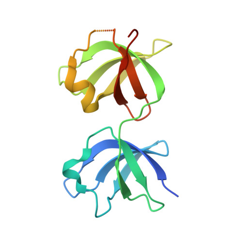
    The proteasome forms the core of the protein quality control system in archaea and eukaryotes and also occurs in one bacterial lineage, the Actinobacteria. Access to its proteolytic compartment is controlled by AAA ATPases, whose N-terminal domains (N domains) are thought to mediate substrate recognition. The N domains of an archaeal proteasomal ATPase, Archaeoglobus fulgidus PAN, and of its actinobacterial homolog, Rhodococcus erythropolis ARC, form hexameric rings, whose subunits consist of an N-terminal coiled coil and a C-terminal OB domain. In ARC-N, the OB domains are duplicated and form separate rings. PAN-N and ARC-N can act as chaperones, preventing the aggregation of heterologous proteins in vitro, and this activity is preserved in various chimeras, even when these include coiled coils and OB domains from unrelated proteins. The structures suggest a molecular mechanism for substrate processing based on concerted radial motions of the coiled coils relative to the OB rings.


  • Organizational Affiliation

    Department of Protein Evolution, Max Planck Institute for Developmental Biology, D-72076 Tübingen, Germany.


Macromolecules
Find similar proteins by:  (by identity cutoff)  |  3D Structure
Entity ID: 1
MoleculeChains Sequence LengthOrganismDetailsImage
ARC
A, B, C
153Rhodococcus erythropolisMutation(s): 0 
UniProt
Find proteins for O50202 (Rhodococcus erythropolis)
Explore O50202 
Go to UniProtKB:  O50202
Entity Groups  
Sequence Clusters30% Identity50% Identity70% Identity90% Identity95% Identity100% Identity
UniProt GroupO50202
Sequence Annotations
Expand
  • Reference Sequence
Experimental Data & Validation

Experimental Data

  • Method: X-RAY DIFFRACTION
  • Resolution: 1.60 Å
  • R-Value Free: 0.246 
  • R-Value Work: 0.208 
  • R-Value Observed: 0.210 
  • Space Group: P 32 2 1
Unit Cell:
Length ( Å )Angle ( ˚ )
a = 100.33α = 90
b = 100.33β = 90
c = 88γ = 120
Software Package:
Software NamePurpose
REFMACrefinement
XDSdata reduction
XDSdata scaling
SHARPphasing

Structure Validation

View Full Validation Report


Entry History 

Deposition Data

Revision History  (Full details and data files)

  • Version 1.0: 2009-05-12
    Type: Initial release
  • Version 1.1: 2011-07-13
    Changes: Advisory, Version format compliance