2W8B

Crystal structure of processed TolB in complex with Pal


Experimental Data Snapshot

  • Method: X-RAY DIFFRACTION
  • Resolution: 1.86 Å
  • R-Value Free: 0.247 
  • R-Value Work: 0.191 
  • R-Value Observed: 0.194 

wwPDB Validation   3D Report Full Report


This is version 1.3 of the entry. See complete history


Literature

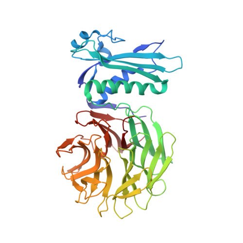
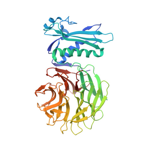
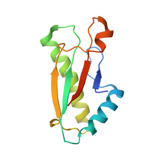
Allosteric Beta-Propeller Signalling in Tolb and its Manipulation by Translocating Colicins.

Bonsor, D.A.Hecht, O.Vankemmelbeke, M.Sharma, A.Krachler, A.M.Housden, N.G.Lilly, K.J.James, R.Moore, G.R.Kleanthous, C.

(2009) EMBO J 28: 2846

  • DOI: https://doi.org/10.1038/emboj.2009.224
  • Primary Citation of Related Structures:  
    2W8B

  • PubMed Abstract: 

    The Tol system is a five-protein assembly parasitized by colicins and bacteriophages that helps stabilize the Gram-negative outer membrane (OM). We show that allosteric signalling through the six-bladed beta-propeller protein TolB is central to Tol function in Escherichia coli and that this is subverted by colicins such as ColE9 to initiate their OM translocation. Protein-protein interactions with the TolB beta-propeller govern two conformational states that are adopted by the distal N-terminal 12 residues of TolB that bind TolA in the inner membrane. ColE9 promotes disorder of this 'TolA box' and recruitment of TolA. In contrast to ColE9, binding of the OM lipoprotein Pal to the same site induces conformational changes that sequester the TolA box to the TolB surface in which it exhibits little or no TolA binding. Our data suggest that Pal is an OFF switch for the Tol assembly, whereas colicins promote an ON state even though mimicking Pal. Comparison of the TolB mechanism to that of vertebrate guanine nucleotide exchange factor RCC1 suggests that allosteric signalling may be more prevalent in beta-propeller proteins than currently realized.


  • Organizational Affiliation

    Department of Biology, University of York, York, UK.


Macromolecules
Find similar proteins by:  (by identity cutoff)  |  3D Structure
Entity ID: 1
MoleculeChains Sequence LengthOrganismDetailsImage
PROTEIN TOLB409Escherichia coliMutation(s): 0 
UniProt
Find proteins for P0A855 (Escherichia coli (strain K12))
Explore P0A855 
Go to UniProtKB:  P0A855
Entity Groups  
Sequence Clusters30% Identity50% Identity70% Identity90% Identity95% Identity100% Identity
UniProt GroupP0A855
Sequence Annotations
Expand
  • Reference Sequence
Find similar proteins by:  (by identity cutoff)  |  3D Structure
Entity ID: 2
MoleculeChains Sequence LengthOrganismDetailsImage
PROTEIN TOLB
B, D, F
409Escherichia coliMutation(s): 0 
UniProt
Find proteins for P0A855 (Escherichia coli (strain K12))
Explore P0A855 
Go to UniProtKB:  P0A855
Entity Groups  
Sequence Clusters30% Identity50% Identity70% Identity90% Identity95% Identity100% Identity
UniProt GroupP0A855
Sequence Annotations
Expand
  • Reference Sequence
Find similar proteins by:  (by identity cutoff)  |  3D Structure
Entity ID: 3
MoleculeChains Sequence LengthOrganismDetailsImage
PEPTIDOGLYCAN-ASSOCIATED LIPOPROTEIN
C, E, G, H
118Escherichia coliMutation(s): 0 
UniProt
Find proteins for P0A912 (Escherichia coli (strain K12))
Explore P0A912 
Go to UniProtKB:  P0A912
Entity Groups  
Sequence Clusters30% Identity50% Identity70% Identity90% Identity95% Identity100% Identity
UniProt GroupP0A912
Sequence Annotations
Expand
  • Reference Sequence
Small Molecules
Ligands 3 Unique
IDChains Name / Formula / InChI Key2D Diagram3D Interactions
SO4
Query on SO4

Download Ideal Coordinates CCD File 
I [auth A]
J [auth A]
K [auth A]
N [auth B]
O [auth B]
I [auth A],
J [auth A],
K [auth A],
N [auth B],
O [auth B],
T [auth D],
X [auth F],
Z [auth G]
SULFATE ION
O4 S
QAOWNCQODCNURD-UHFFFAOYSA-L
GOL
Query on GOL

Download Ideal Coordinates CCD File 
L [auth A]
P [auth B]
Q [auth B]
S [auth C]
U [auth D]
L [auth A],
P [auth B],
Q [auth B],
S [auth C],
U [auth D],
V [auth D],
Y [auth F]
GLYCEROL
C3 H8 O3
PEDCQBHIVMGVHV-UHFFFAOYSA-N
ACT
Query on ACT

Download Ideal Coordinates CCD File 
AA [auth H],
M [auth A],
R [auth B],
W [auth D]
ACETATE ION
C2 H3 O2
QTBSBXVTEAMEQO-UHFFFAOYSA-M
Experimental Data & Validation

Experimental Data

  • Method: X-RAY DIFFRACTION
  • Resolution: 1.86 Å
  • R-Value Free: 0.247 
  • R-Value Work: 0.191 
  • R-Value Observed: 0.194 
  • Space Group: P 1
Unit Cell:
Length ( Å )Angle ( ˚ )
a = 74.74α = 86.81
b = 89.24β = 89.81
c = 90.9γ = 68.62
Software Package:
Software NamePurpose
REFMACrefinement
MOSFLMdata reduction
SCALAdata scaling
REFMACphasing

Structure Validation

View Full Validation Report


Entry History 

Deposition Data

Revision History  (Full details and data files)

  • Version 1.0: 2009-02-17
    Type: Initial release
  • Version 1.1: 2011-05-08
    Changes: Version format compliance
  • Version 1.2: 2011-07-13
    Changes: Version format compliance
  • Version 1.3: 2023-12-13
    Changes: Data collection, Database references, Derived calculations, Other, Refinement description