2W86

Crystal structure of fibrillin-1 domains cbEGF9hyb2cbEGF10, calcium saturated form


Experimental Data Snapshot

  • Method: X-RAY DIFFRACTION
  • Resolution: 1.80 Å
  • R-Value Free: 0.221 
  • R-Value Work: 0.185 
  • R-Value Observed: 0.187 

wwPDB Validation   3D Report Full Report


This is version 1.2 of the entry. See complete history


Literature

Structure and Interdomain Interactions of a Hybrid Domain: A Disulphide-Rich Module of the Fibrillin/Ltbp Superfamily of Matrix Proteins.

Jensen, S.A.Iqbal, S.Lowe, E.D.Redfield, C.Handford, P.A.

(2009) Structure 17: 759

  • DOI: https://doi.org/10.1016/j.str.2009.03.014
  • Primary Citation of Related Structures:  
    2W86

  • PubMed Abstract: 

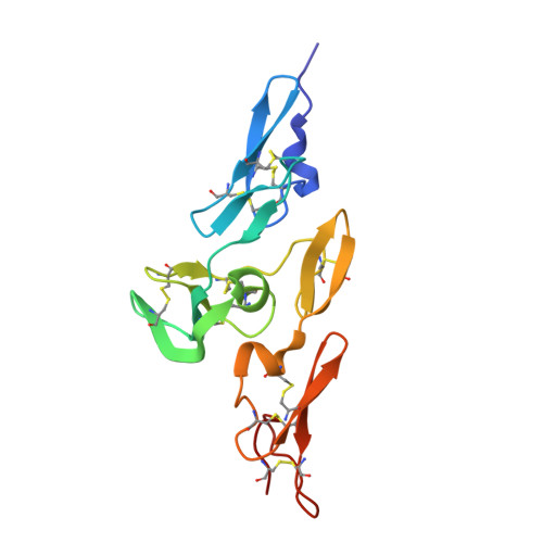
    The fibrillins and latent transforming growth factor-beta binding proteins (LTBPs) form a superfamily of structurally-related proteins consisting of calcium-binding epidermal growth factor-like (cbEGF) domains interspersed with 8-cysteine-containing transforming growth factor beta-binding protein-like (TB) and hybrid (hyb) domains. Fibrillins are the major components of the extracellular 10-12 nm diameter microfibrils, which mediate a variety of cell-matrix interactions. Here we present the crystal structure of a fibrillin-1 cbEGF9-hyb2-cbEGF10 fragment, solved to 1.8 A resolution. The hybrid domain fold is similar, but not identical, to the TB domain fold seen in previous fibrillin-1 and LTBP-1 fragments. Pairwise interactions with neighboring cbEGF domains demonstrate extensive interfaces, with the hyb2-cbEGF10 interface dependent on Ca(2+) binding. These observations provide accurate constraints for models of fibrillin organization within the 10-12 nm microfibrils and provide further molecular insights into how Ca(2+) binding influences the intermolecular interactions and biomechanical properties of fibrillin-1.


  • Organizational Affiliation

    Department of Biochemistry, University of Oxford, Oxford, UK.


Macromolecules
Find similar proteins by:  (by identity cutoff)  |  3D Structure
Entity ID: 1
MoleculeChains Sequence LengthOrganismDetailsImage
FIBRILLIN-1147Homo sapiensMutation(s): 0 
UniProt & NIH Common Fund Data Resources
Find proteins for P35555 (Homo sapiens)
Explore P35555 
Go to UniProtKB:  P35555
PHAROS:  P35555
GTEx:  ENSG00000166147 
Entity Groups  
Sequence Clusters30% Identity50% Identity70% Identity90% Identity95% Identity100% Identity
UniProt GroupP35555
Sequence Annotations
Expand
  • Reference Sequence
Experimental Data & Validation

Experimental Data

  • Method: X-RAY DIFFRACTION
  • Resolution: 1.80 Å
  • R-Value Free: 0.221 
  • R-Value Work: 0.185 
  • R-Value Observed: 0.187 
  • Space Group: P 21 21 21
Unit Cell:
Length ( Å )Angle ( ˚ )
a = 31.232α = 90
b = 47.258β = 90
c = 89.105γ = 90
Software Package:
Software NamePurpose
MOSFLMdata reduction
SCALAdata scaling
SHELXDphasing
SHARPphasing
PHENIXrefinement

Structure Validation

View Full Validation Report


Entry History 

Deposition Data

Revision History  (Full details and data files)

  • Version 1.0: 2009-05-26
    Type: Initial release
  • Version 1.1: 2011-05-08
    Changes: Version format compliance
  • Version 1.2: 2011-07-13
    Changes: Version format compliance