2W3O

Crystal structure of the human PNKP FHA domain in complex with an XRCC1-derived phosphopeptide


Experimental Data Snapshot

  • Method: X-RAY DIFFRACTION
  • Resolution: 1.85 Å
  • R-Value Free: 0.236 
  • R-Value Work: 0.178 
  • R-Value Observed: 0.181 

wwPDB Validation   3D Report Full Report


This is version 1.3 of the entry. See complete history


Literature

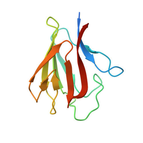
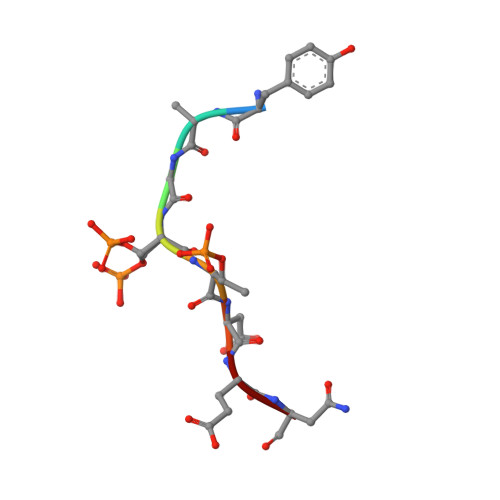
Specific Recognition of a Multiply Phosphorylated Motif in the DNA Repair Scaffold Xrcc1 by the Fha Domain of Human Pnk.

Ali, A.A.E.Jukes, R.M.Pearl, L.H.Oliver, A.W.

(2009) Nucleic Acids Res 37: 1701

  • DOI: https://doi.org/10.1093/nar/gkn1086
  • Primary Citation of Related Structures:  
    2BRF, 2W3O

  • PubMed Abstract: 

    Short-patch repair of DNA single-strand breaks and gaps (SSB) is coordinated by XRCC1, a scaffold protein that recruits the DNA polymerase and DNA ligase required for filling and sealing the damaged strand. XRCC1 can also recruit end-processing enzymes, such as PNK (polynucleotide kinase 3'-phosphatase), Aprataxin and APLF (aprataxin/PNK-like factor), which ensure the availability of a free 3'-hydroxyl on one side of the gap, and a 5'-phosphate group on the other, for the polymerase and ligase reactions respectively. PNK binds to a phosphorylated segment of XRCC1 (between its two C-terminal BRCT domains) via its Forkhead-associated (FHA) domain. We show here, contrary to previous studies, that the FHA domain of PNK binds specifically, and with high affinity to a multiply phosphorylated motif in XRCC1 containing a pSer-pThr dipeptide, and forms a 2:1 PNK:XRCC1 complex. The high-resolution crystal structure of a PNK-FHA-XRCC1 phosphopeptide complex reveals the basis for this unusual bis-phosphopeptide recognition, which is probably a common feature of the known XRCC1-associating end-processing enzymes.


  • Organizational Affiliation

    Cancer Research UK DNA Repair Enzyme Group, Section of Structural Biology, The Institute of Cancer Research, London SW3 6JB, UK.


Macromolecules
Find similar proteins by:  (by identity cutoff)  |  3D Structure
Entity ID: 1
MoleculeChains Sequence LengthOrganismDetailsImage
BIFUNCTIONAL POLYNUCLEOTIDE PHOSPHATASE/KINASE
A, B
113Homo sapiensMutation(s): 1 
EC: 3.1.3.32 (PDB Primary Data), 2.7.1.78 (PDB Primary Data)
UniProt & NIH Common Fund Data Resources
Find proteins for Q96T60 (Homo sapiens)
Explore Q96T60 
Go to UniProtKB:  Q96T60
PHAROS:  Q96T60
GTEx:  ENSG00000039650 
Entity Groups  
Sequence Clusters30% Identity50% Identity70% Identity90% Identity95% Identity100% Identity
UniProt GroupQ96T60
Sequence Annotations
Expand
  • Reference Sequence

Find similar proteins by:  Sequence   |   3D Structure  

Entity ID: 2
MoleculeChains Sequence LengthOrganismDetailsImage
DNA REPAIR PROTEIN XRCC1
C, D
8Homo sapiensMutation(s): 0 
UniProt & NIH Common Fund Data Resources
Find proteins for P18887 (Homo sapiens)
Explore P18887 
Go to UniProtKB:  P18887
PHAROS:  P18887
GTEx:  ENSG00000073050 
Entity Groups  
UniProt GroupP18887
Sequence Annotations
Expand
  • Reference Sequence
Experimental Data & Validation

Experimental Data

  • Method: X-RAY DIFFRACTION
  • Resolution: 1.85 Å
  • R-Value Free: 0.236 
  • R-Value Work: 0.178 
  • R-Value Observed: 0.181 
  • Space Group: P 3
Unit Cell:
Length ( Å )Angle ( ˚ )
a = 56.9α = 90
b = 56.9β = 90
c = 62.77γ = 120
Software Package:
Software NamePurpose
REFMACrefinement
MOSFLMdata reduction
SCALAdata scaling
PHASERphasing

Structure Validation

View Full Validation Report


Entry History 

Deposition Data

Revision History  (Full details and data files)

  • Version 1.0: 2009-02-03
    Type: Initial release
  • Version 1.1: 2011-05-08
    Changes: Version format compliance
  • Version 1.2: 2011-07-13
    Changes: Version format compliance
  • Version 1.3: 2023-12-13
    Changes: Data collection, Database references, Derived calculations, Other, Refinement description