2.3 A X-Ray Structure of the Heme-Bound Gaf Domain of Sensory Histidine Kinase Dost of Mycobacterium Tuberculosis.
Podust, L.M., Ioanoviciu, A., Ortiz de Montellano, P.R.(2008) Biochemistry 47: 12523
- PubMed: 18980385
- DOI: https://doi.org/10.1021/bi8012356
- Primary Citation of Related Structures:
2VZW - PubMed Abstract:
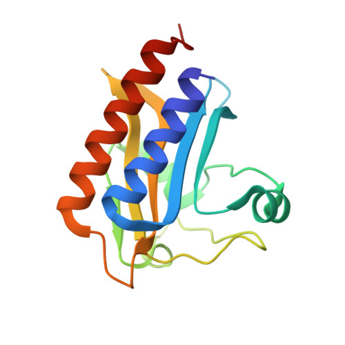
Mycobacterium tuberculosis responds to changes in environmental conditions through a two-component signaling system that detects reduced O(2) tension and NO and CO exposures via the heme-binding GAF domains of two sensory histidine kinases, DosT and DevS, and the transcriptional regulator DosR. We report the first X-ray structure of the DosT heme-bound GAF domain (GAF(DosT)) in both oxy and deoxy forms determined to a resolution of 2.3 A. In GAF(DosT), heme binds in an orientation orthogonal to that in the PAS domains via a highly conserved motif, including invariant H147 as a proximal heme axial ligand. On the distal side, invariant Y169 forms stacking interactions with the heme with its long axis parallel and the plane of the ring orthogonal to the heme plane. In one of the two protein monomers in an asymmetric unit, O(2) binds as a second axial ligand to the heme iron and is stabilized via a H-bond to the OH group of Y169. The structure reveals two small tunnel-connected cavities and a pore on the protein surface that suggest a potential route for the access of O(2) to the sensing pocket. The limited conformational differences observed between differently heme iron-ligated GAF(DosT) monomers in the asymmetric unit may result from crystal lattice limitations since atmospheric oxygen binding likely occurs in the crystal as a result of X-ray-induced Fe(3+) photoreduction during diffraction data collection. Determination of the GAF(DosT) structure sets up a framework in which to address ligand recognition, discrimination, and signal propagation schemes in the heme-based GAF domains of biological sensors.
Organizational Affiliation:
Department of Pharmaceutical Chemistry, University of California, 600 16th Street, San Francisco, California 94158-2517, USA.