D-Ribose-5-Phosphate Isomerase B from Escherichia Coli is Also a Functional D-Allose-6-Phosphate Isomerase, While the Mycobacterium Tuberculosis Enzyme is not.
Roos, A.K., Mariano, S., Kowalinski, E., Salmon, L., Mowbray, S.L.(2008) J Mol Biol 382: 667
- PubMed: 18640127
- DOI: https://doi.org/10.1016/j.jmb.2008.06.090
- Primary Citation of Related Structures:
2VVO, 2VVP, 2VVQ, 2VVR - PubMed Abstract:
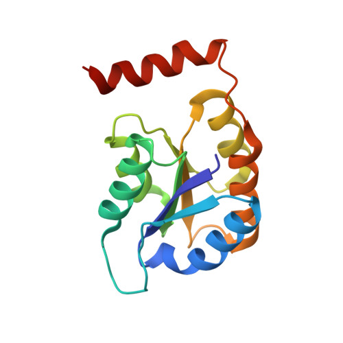
Interconversion of D-ribose-5-phosphate (R5P) and D-ribulose-5-phosphate is an important step in the pentose phosphate pathway. Two unrelated enzymes with R5P isomerase activity were first identified in Escherichia coli, RpiA and RpiB. In this organism, the essential 5-carbon sugars were thought to be processed by RpiA, while the primary role of RpiB was suggested to instead be interconversion of the rare 6-carbon sugars D-allose-6-phosphate (All6P) and D-allulose-6-phosphate. In Mycobacterium tuberculosis, where only an RpiB is found, the 5-carbon sugars are believed to be the enzyme's primary substrates. Here, we present kinetic studies examining the All6P isomerase activity of the RpiBs from these two organisms and show that only the E. coli enzyme can catalyze the reaction efficiently. All6P instead acts as an inhibitor of the M. tuberculosis enzyme in its action on R5P. X-ray studies of the M. tuberculosis enzyme co-crystallized with All6P and 5-deoxy-5-phospho-D-ribonohydroxamate (an inhibitor designed to mimic the 6-carbon sugar) and comparison with the E. coli enzyme's structure allowed us to identify differences in the active sites that explain the kinetic results. Two other structures, that of a mutant E. coli RpiB in which histidine 99 was changed to asparagine and that of wild-type M. tuberculosis enzyme, both co-crystallized with the substrate ribose-5-phosphate, shed additional light on the reaction mechanism of RpiBs generally.
Organizational Affiliation:
Department of Cell and Molecular Biology, Biomedical Center, Uppsala University, Box 596, SE-751 24 Uppsala, Sweden.