Ordering of C-Terminal Loop and Glutaminase Domains of Glucosamine-6-Phosphate Synthase Promotes Sugar Ring Opening and Formation of the Ammonia Channel.
Mouilleron, S., Badet-Denisot, M.-A., Golinelli-Pimpaneau, B.(2008) J Mol Biol 377: 1174
- PubMed: 18295797
- DOI: https://doi.org/10.1016/j.jmb.2008.01.077
- Primary Citation of Related Structures:
2VF4 - PubMed Abstract:
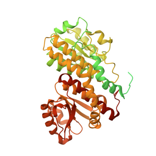
Glucosamine-6-phosphate synthase (GlmS) channels ammonia from glutamine at the glutaminase site to fructose 6-phosphate (Fru6P) at the synthase site. Escherichia coli GlmS is composed of two C-terminal synthase domains that form the dimer interface and two N-terminal glutaminase domains at its periphery. We report the crystal structures of GlmS alone and in complex with the glucosamine-6-phosphate product at 2.95 A and 2.9 A resolution, respectively. Surprisingly, although the whole protein is present in this crystal form, no electron density for the glutaminase domain was observed, indicating its mobility. Comparison of the two structures with that of the previously reported GlmS-Fru6P complex shows that, upon sugar binding, the C-terminal loop, which forms the major part of the channel walls, becomes ordered and covers the synthase site. The ordering of the glutaminase domains likely follows Fru6P binding by the anchoring of Trp74, which acts as the gate of the channel, on the closed C-terminal loop. This is accompanied by a major conformational change of the side chain of Lys503# of the neighboring synthase domain that strengthens the interactions of the synthase domain with the C-terminal loop and completely shields the synthase site. The concomitant conformational change of the Lys503#-Gly505# tripeptide places catalytic His504# in the proper position to open the sugar and buries the linear sugar, which is now in the vicinity of the catalytic groups involved in the sugar isomerization reaction. Together with the previously reported structures of GlmS in complex with Fru6P or glucose 6-phosphate and a glutamine analogue, the new structures reveal the structural changes occurring during the whole catalytic cycle.
Organizational Affiliation:
Laboratoire d'Enzymologie et Biochimie Structurales, Bâtiment 34. CNRS, 1 avenue de la Terrasse, 91190 Gif-sur-Yvette, France.