2V7N

Unusual twinning in crystals of the CitS binding antibody Fab fragment f3p4


Experimental Data Snapshot

  • Method: X-RAY DIFFRACTION
  • Resolution: 1.92 Å
  • R-Value Free: 0.202 
  • R-Value Observed: 0.155 

wwPDB Validation   3D Report Full Report


This is version 1.2 of the entry. See complete history


Literature

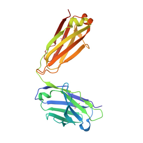
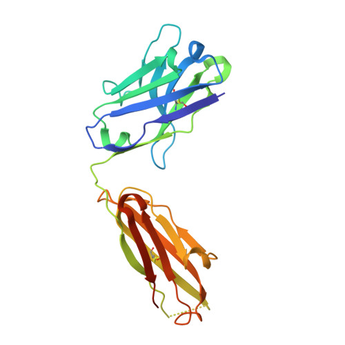
Structure of the Recombinant Antibody Fab Fragment F3P4.

Frey, D.Huber, T.Plueckthun, A.Gruetter, M.G.

(2008) Acta Crystallogr D Biol Crystallogr 64: 636

  • DOI: https://doi.org/10.1107/S0907444908007282
  • Primary Citation of Related Structures:  
    2V7N

  • PubMed Abstract: 

    The structure of the antibody Fab fragment f3p4, which was selected from a subset of the synthetic HuCAL antibody library to bind the sodium citrate symporter CitS, is described at 1.92 A resolution. Comparison with computational models revealed deviations in a few framework positions and in the binding loops. The crystals belong to space group P2(1)2(1)2 and contain four molecules in the asymmetric unit, with unit-cell parameters a=102.77, b=185.92, c=102.97 A. These particular unit-cell parameters allowed pseudo-merohedral twinning; interestingly, the twinning law relates a twofold screw axis to a twofold axis.


  • Organizational Affiliation

    Department of Biochemistry, University of Zürich, Switzerland.


Macromolecules
Find similar proteins by:  (by identity cutoff)  |  3D Structure
Entity ID: 1
MoleculeChains Sequence LengthOrganismDetailsImage
IMMUNOGLOBULIN LIGHT CHAIN
A, C, E, G
215synthetic constructMutation(s): 0 
Entity Groups  
Sequence Clusters30% Identity50% Identity70% Identity90% Identity95% Identity100% Identity
Sequence Annotations
Expand
  • Reference Sequence
Find similar proteins by:  (by identity cutoff)  |  3D Structure
Entity ID: 2
MoleculeChains Sequence LengthOrganismDetailsImage
IMMUNOGLOBULIN HEAVY CHAIN
B, D, F, H
229synthetic constructMutation(s): 0 
Entity Groups  
Sequence Clusters30% Identity50% Identity70% Identity90% Identity95% Identity100% Identity
Sequence Annotations
Expand
  • Reference Sequence
Experimental Data & Validation

Experimental Data

  • Method: X-RAY DIFFRACTION
  • Resolution: 1.92 Å
  • R-Value Free: 0.202 
  • R-Value Observed: 0.155 
  • Space Group: P 21 21 2
Unit Cell:
Length ( Å )Angle ( ˚ )
a = 102.79α = 90
b = 185.92β = 90
c = 102.99γ = 90
Software Package:
Software NamePurpose
SHELXL-97refinement
XDSdata reduction
XDSdata scaling
PHASERphasing

Structure Validation

View Full Validation Report


Entry History 

Deposition Data

Revision History  (Full details and data files)

  • Version 1.0: 2008-06-17
    Type: Initial release
  • Version 1.1: 2012-05-30
    Changes: Database references, Derived calculations, Non-polymer description, Other, Source and taxonomy, Version format compliance
  • Version 1.2: 2019-05-22
    Changes: Data collection, Other, Refinement description