2V53

Crystal structure of a SPARC-collagen complex


Experimental Data Snapshot

  • Method: X-RAY DIFFRACTION
  • Resolution: 3.20 Å
  • R-Value Free: 0.320 
  • R-Value Work: 0.254 
  • R-Value Observed: 0.261 

wwPDB Validation   3D Report Full Report


This is version 2.1 of the entry. See complete history


Literature

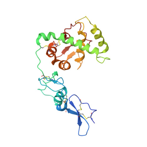
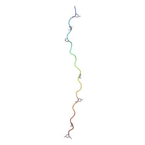
Structural Basis of Sequence-Specific Collagen Recognition by Sparc.

Hohenester, E.Sasaki, T.Giudici, C.Farndale, R.W.Bachinger, H.P.

(2008) Proc Natl Acad Sci U S A 105: 18273

  • DOI: https://doi.org/10.1073/pnas.0808452105
  • Primary Citation of Related Structures:  
    2V53

  • PubMed Abstract: 

    Protein interactions with the collagen triple helix play a critical role in collagen fibril formation, cell adhesion, and signaling. However, structural insight into sequence-specific collagen recognition is limited to an integrin-peptide complex. A GVMGFO motif in fibrillar collagens (O denotes 4-hydroxyproline) binds 3 unrelated proteins: von Willebrand factor (VWF), discoidin domain receptor 2 (DDR2), and the extracellular matrix protein SPARC/osteonectin/BM-40. We report the crystal structure at 3.2 A resolution of human SPARC bound to a triple-helical 33-residue peptide harboring the promiscuous GVMGFO motif. SPARC recognizes the GVMGFO motifs of the middle and trailing collagen chains, burying a total of 720 A(2) of solvent-accessible collagen surface. SPARC binding does not distort the canonical triple helix of the collagen peptide. In contrast, a critical loop in SPARC is substantially remodelled upon collagen binding, creating a deep pocket that accommodates the phenylalanine residue of the trailing collagen chain ("Phe pocket"). This highly restrictive specificity pocket is shared with the collagen-binding integrin I-domains but differs strikingly from the shallow collagen-binding grooves of the platelet receptor glycoprotein VI and microbial adhesins. We speculate that binding of the GVMGFO motif to VWF and DDR2 also results in structural changes and the formation of a Phe pocket.


  • Organizational Affiliation

    Department of Life Sciences, Imperial College London, London SW7 2AZ, United Kingdom. e.hohenester@imperial.ac.uk


Macromolecules
Find similar proteins by:  (by identity cutoff)  |  3D Structure
Entity ID: 1
MoleculeChains Sequence LengthOrganismDetailsImage
SPARC230Homo sapiensMutation(s): 0 
Membrane Entity: Yes 
UniProt & NIH Common Fund Data Resources
Find proteins for P09486 (Homo sapiens)
Explore P09486 
Go to UniProtKB:  P09486
PHAROS:  P09486
GTEx:  ENSG00000113140 
Entity Groups  
Sequence Clusters30% Identity50% Identity70% Identity90% Identity95% Identity100% Identity
UniProt GroupP09486
Sequence Annotations
Expand
  • Reference Sequence
Find similar proteins by:  (by identity cutoff)  |  3D Structure
Entity ID: 2
MoleculeChains Sequence LengthOrganismDetailsImage
COLLAGEN ALPHA-1(III) CHAIN
B, C, D
33Homo sapiensMutation(s): 0 
Membrane Entity: Yes 
UniProt & NIH Common Fund Data Resources
Find proteins for P02461 (Homo sapiens)
Explore P02461 
Go to UniProtKB:  P02461
PHAROS:  P02461
GTEx:  ENSG00000168542 
Entity Groups  
Sequence Clusters30% Identity50% Identity70% Identity90% Identity95% Identity100% Identity
UniProt GroupP02461
Sequence Annotations
Expand
  • Reference Sequence
Oligosaccharides

Help

Entity ID: 3
MoleculeChains Length2D Diagram Glycosylation3D Interactions
2-acetamido-2-deoxy-beta-D-glucopyranose-(1-4)-2-acetamido-2-deoxy-beta-D-glucopyranose
E
2N-Glycosylation
Glycosylation Resources
GlyTouCan:  G42666HT
GlyCosmos:  G42666HT
GlyGen:  G42666HT
Small Molecules
Modified Residues  1 Unique
IDChains TypeFormula2D DiagramParent
HYP
Query on HYP
B, C, D
L-PEPTIDE LINKINGC5 H9 N O3PRO
Experimental Data & Validation

Experimental Data

  • Method: X-RAY DIFFRACTION
  • Resolution: 3.20 Å
  • R-Value Free: 0.320 
  • R-Value Work: 0.254 
  • R-Value Observed: 0.261 
  • Space Group: P 42 21 2
Unit Cell:
Length ( Å )Angle ( ˚ )
a = 89.79α = 90
b = 89.79β = 90
c = 127.1γ = 90
Software Package:
Software NamePurpose
MOSFLMdata reduction
SCALAdata scaling
PHASERphasing
autoSHARPphasing
PHENIXrefinement

Structure Validation

View Full Validation Report


Entry History 

Deposition Data

Revision History  (Full details and data files)

  • Version 1.0: 2008-11-25
    Type: Initial release
  • Version 1.1: 2011-05-08
    Changes: Version format compliance
  • Version 1.2: 2011-07-13
    Changes: Version format compliance
  • Version 2.0: 2020-07-29
    Type: Remediation
    Reason: Carbohydrate remediation
    Changes: Atomic model, Data collection, Derived calculations, Other, Structure summary
  • Version 2.1: 2023-12-13
    Changes: Data collection, Database references, Refinement description, Structure summary