Molecular dynamics refinement of a thermitase-eglin-c complex at 1.98 A resolution and comparison of two crystal forms that differ in calcium content.
Gros, P., Betzel, C., Dauter, Z., Wilson, K.S., Hol, W.G.(1989) J Mol Biol 210: 347-367
- PubMed: 2689655
- DOI: https://doi.org/10.1016/0022-2836(89)90336-7
- Primary Citation of Related Structures:
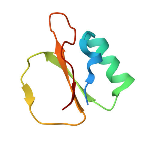
2TEC - PubMed Abstract:
The crystal structure of the complex of thermitase with eglin-c in crystal form II, obtained in the presence of 5 mM-CaCl2, has been determined at 1.98 A resolution. The structure was solved by a molecular replacement method, then molecular dynamics crystallographic refinement was started using the thermitase-eglin-c structure as determined for crystal form I. A ten degrees rigid body misplacement of the core of eglin-c was corrected by the molecular dynamics crystallographic refinement without any need for manual rebuilding on a graphics system. A final crystallographic R-factor of 16.5% was obtained for crystal form II. The comparison of the complexes of thermitase with eglin-c in the two crystal forms shows that the eglin-c cores are differently oriented with respect to the protease. The inhibiting loop of eglin binds in a similar way to thermitase as to subtilisin Carlsberg. A tryptophanyl residue at the S4 site explains the preference of thermitase for aromatic residues of the substrate at the P4 site. The difference in the P1 binding pocket, asparagine in thermitase instead of glycine in subtilisin Carlsberg, does not change the binding of eglin-c. The preference for an arginyl residue at the P1 site of thermitase can be explained by the hydrogen bonding with Asn170 in thermitase. Three ion-binding sites of thermitase have been identified. The strong and weak calcium-binding sites resemble the equivalent sites of subtilisin Carlsberg and subtilisin BPN', though there are important amino acid differences at the calcium-binding sites. The medium-strength calcium-binding site of thermitase is observed in the subtilisin family for the first time. The calcium is bound to residues from the loop 57 to 66. A difference in chelation is observed at this site between the two crystal forms of thermitase, which differ in calcium concentration. Additional electron density is observed near Asp60 in crystal form II, which has more calcium bound than form I. This density is possibly due to a water molecule ligating the calcium ion or the result of Asp60 assuming two significantly different conformations.
Organizational Affiliation:
Laboratory of Chemical Physics, University of Groningen, The Netherlands.