2RDL

Hamster Chymase 2


Experimental Data Snapshot

  • Method: X-RAY DIFFRACTION
  • Resolution: 2.50 Å
  • R-Value Free: 0.253 
  • R-Value Work: 0.186 

wwPDB Validation   3D Report Full Report


This is version 1.2 of the entry. See complete history


Literature

Structural basis for elastolytic substrate specificity in rodent alpha-chymases.

Kervinen, J.Abad, M.Crysler, C.Kolpak, M.Mahan, A.D.Masucci, J.A.Bayoumy, S.Cummings, M.D.Yao, X.Olson, M.de Garavilla, L.Kuo, L.Deckman, I.Spurlino, J.

(2008) J Biol Chem 283: 427-436

  • DOI: https://doi.org/10.1074/jbc.M707157200
  • Primary Citation of Related Structures:  
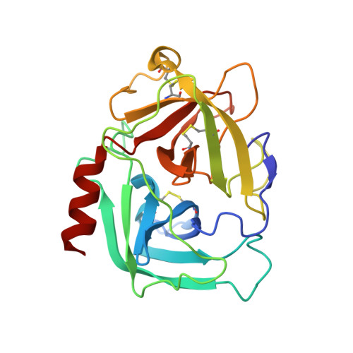
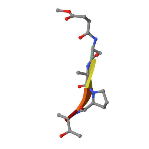
    2RDL

  • PubMed Abstract: 

    Divergence of substrate specificity within the context of a common structural framework represents an important mechanism by which new enzyme activity naturally evolves. We present enzymological and x-ray structural data for hamster chymase-2 (HAM2) that provides a detailed explanation for the unusual hydrolytic specificity of this rodent alpha-chymase. In enzymatic characterization, hamster chymase-1 (HAM1) showed typical chymase proteolytic activity. In contrast, HAM2 exhibited atypical substrate specificity, cleaving on the carboxyl side of the P1 substrate residues Ala and Val, characteristic of elastolytic rather than chymotryptic specificity. The 2.5-A resolution crystal structure of HAM2 complexed to the peptidyl inhibitor MeOSuc-Ala-Ala-Pro-Ala-chloromethylketone revealed a narrow and shallow S1 substrate binding pocket that accommodated only a small hydrophobic residue (e.g. Ala or Val). The different substrate specificities of HAM2 and HAM1 are explained by changes in four S1 substrate site residues (positions 189, 190, 216, and 226). Of these, Asn(189), Val(190), and Val(216) form an easily identifiable triplet in all known rodent alpha-chymases that can be used to predict elastolytic specificity for novel chymase-like sequences. Phylogenetic comparison defines guinea pig and rabbit chymases as the closest orthologs to rodent alpha-chymases.


  • Organizational Affiliation

    Johnson & Johnson Pharmaceutical Research and Development, Structural Biology, Exton, Pennsylvania 19341. Electronic address: jkervine@prdus.jnj.com.


Macromolecules
Find similar proteins by:  (by identity cutoff)  |  3D Structure
Entity ID: 1
MoleculeChains Sequence LengthOrganismDetailsImage
Chymase 2
A, B
226Mesocricetus auratusMutation(s): 0 
UniProt
Find proteins for O70164 (Mesocricetus auratus)
Explore O70164 
Go to UniProtKB:  O70164
Entity Groups  
Sequence Clusters30% Identity50% Identity70% Identity90% Identity95% Identity100% Identity
UniProt GroupO70164
Sequence Annotations
Expand
  • Reference Sequence

Find similar proteins by:  Sequence   |   3D Structure  

Entity ID: 2
MoleculeChains Sequence LengthOrganismDetailsImage
METHOXYSUCCINYL-ALA-ALA-PRO-ALA-CHLOROMETHYLKETONE INHIBITORC [auth I],
D [auth J]
6N/AMutation(s): 0 
Sequence Annotations
Expand
  • Reference Sequence
Small Molecules
Modified Residues  1 Unique
IDChains TypeFormula2D DiagramParent
ALV
Query on ALV
C [auth I],
D [auth J]
PEPTIDE-LIKEC3 H9 N O2ALA
Biologically Interesting Molecules (External Reference) 1 Unique
Experimental Data & Validation

Experimental Data

  • Method: X-RAY DIFFRACTION
  • Resolution: 2.50 Å
  • R-Value Free: 0.253 
  • R-Value Work: 0.186 
  • Space Group: P 43 21 2
Unit Cell:
Length ( Å )Angle ( ˚ )
a = 71.272α = 90
b = 71.272β = 90
c = 198.575γ = 90
Software Package:
Software NamePurpose
PHENIXrefinement
PDB_EXTRACTdata extraction
HKL-2000data collection
HKL-2000data reduction
HKL-2000data scaling

Structure Validation

View Full Validation Report


Entry History 

Deposition Data

Revision History  (Full details and data files)

  • Version 1.0: 2007-10-30
    Type: Initial release
  • Version 1.1: 2011-07-13
    Changes: Atomic model, Database references, Derived calculations, Non-polymer description, Structure summary, Version format compliance
  • Version 1.2: 2012-12-12
    Changes: Other