2QYF

Crystal structure of the Mad2/p31(comet)/Mad2-binding peptide ternary complex


Experimental Data Snapshot

  • Method: X-RAY DIFFRACTION
  • Resolution: 2.30 Å
  • R-Value Free: 0.257 
  • R-Value Work: 0.187 
  • R-Value Observed: 0.190 

wwPDB Validation   3D Report Full Report


This is version 1.3 of the entry. See complete history


Literature

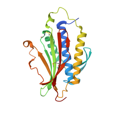
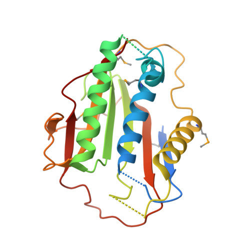
p31comet blocks Mad2 activation through structural mimicry.

Yang, M.Li, B.Tomchick, D.R.Machius, M.Rizo, J.Yu, H.Luo, X.

(2007) Cell 131: 744-755

  • DOI: https://doi.org/10.1016/j.cell.2007.08.048
  • Primary Citation of Related Structures:  
    2QYF

  • PubMed Abstract: 

    The status of spindle checkpoint signaling depends on the balance of two opposing dynamic processes that regulate the highly unusual two-state behavior of Mad2. In mitosis, a Mad1-Mad2 core complex recruits cytosolic Mad2 to kinetochores through Mad2 dimerization and converts Mad2 to a conformer amenable to Cdc20 binding, thereby facilitating checkpoint activation. p31(comet) inactivates the checkpoint through binding to Mad1- or Cdc20-bound Mad2, thereby preventing Mad2 activation and promoting the dissociation of the Mad2-Cdc20 complex. Here, we report the crystal structure of the Mad2-p31(comet) complex. The C-terminal region of Mad2 that undergoes rearrangement in different Mad2 conformers is a major structural determinant for p31(comet) binding, explaining the specificity of p31(comet) toward Mad1- or Cdc20-bound Mad2. p31(comet) adopts a fold strikingly similar to that of Mad2 and binds at the dimerization interface of Mad2. Thus, p31(comet) exploits the two-state behavior of Mad2 to block its activation by acting as an "anti-Mad2."


  • Organizational Affiliation

    Department of Pharmacology, The University of Texas Southwestern Medical Center, 6001 Forest Park Road, Dallas, TX 75390, USA.


Macromolecules
Find similar proteins by:  (by identity cutoff)  |  3D Structure
Entity ID: 1
MoleculeChains Sequence LengthOrganismDetailsImage
Mitotic spindle assembly checkpoint protein MAD2A
A, C
206Homo sapiensMutation(s): 1 
Gene Names: MAD2L1MAD2
UniProt & NIH Common Fund Data Resources
Find proteins for Q13257 (Homo sapiens)
Explore Q13257 
Go to UniProtKB:  Q13257
PHAROS:  Q13257
GTEx:  ENSG00000164109 
Entity Groups  
Sequence Clusters30% Identity50% Identity70% Identity90% Identity95% Identity100% Identity
UniProt GroupQ13257
Sequence Annotations
Expand
  • Reference Sequence
Find similar proteins by:  (by identity cutoff)  |  3D Structure
Entity ID: 2
MoleculeChains Sequence LengthOrganismDetailsImage
MAD2L1-binding protein
B, D
240Homo sapiensMutation(s): 0 
Gene Names: MAD2L1BPCMT2KIAA0110
UniProt & NIH Common Fund Data Resources
Find proteins for Q15013 (Homo sapiens)
Explore Q15013 
Go to UniProtKB:  Q15013
PHAROS:  Q15013
GTEx:  ENSG00000124688 
Entity Groups  
Sequence Clusters30% Identity50% Identity70% Identity90% Identity95% Identity100% Identity
UniProt GroupQ15013
Sequence Annotations
Expand
  • Reference Sequence

Find similar proteins by:  Sequence   |   3D Structure  

Entity ID: 3
MoleculeChains Sequence LengthOrganismDetailsImage
peptide
E, F
12N/AMutation(s): 0 
Entity Groups  
Sequence Clusters30% Identity50% Identity70% Identity90% Identity95% Identity100% Identity
Sequence Annotations
Expand
  • Reference Sequence
Small Molecules
Modified Residues  1 Unique
IDChains TypeFormula2D DiagramParent
MSE
Query on MSE
A, C
L-PEPTIDE LINKINGC5 H11 N O2 SeMET
Experimental Data & Validation

Experimental Data

  • Method: X-RAY DIFFRACTION
  • Resolution: 2.30 Å
  • R-Value Free: 0.257 
  • R-Value Work: 0.187 
  • R-Value Observed: 0.190 
  • Space Group: P 21 21 21
Unit Cell:
Length ( Å )Angle ( ˚ )
a = 68.667α = 90
b = 104.519β = 90
c = 138.825γ = 90
Software Package:
Software NamePurpose
REFMACrefinement
HKL-2000data collection
HKL-2000data reduction
HKL-2000data scaling
MLPHAREphasing

Structure Validation

View Full Validation Report


Entry History 

Deposition Data

Revision History  (Full details and data files)

  • Version 1.0: 2008-01-29
    Type: Initial release
  • Version 1.1: 2011-07-13
    Changes: Version format compliance
  • Version 1.2: 2013-09-18
    Changes: Derived calculations
  • Version 1.3: 2021-10-20
    Changes: Database references, Derived calculations