2QXF

Product bound structure of exonuclease I at 1.5 angstrom resolution


Experimental Data Snapshot

  • Method: X-RAY DIFFRACTION
  • Resolution: 1.50 Å
  • R-Value Free: 0.224 
  • R-Value Work: 0.199 
  • R-Value Observed: 0.200 

wwPDB Validation   3D Report Full Report


Ligand Structure Quality Assessment 


This is version 1.3 of the entry. See complete history


Literature

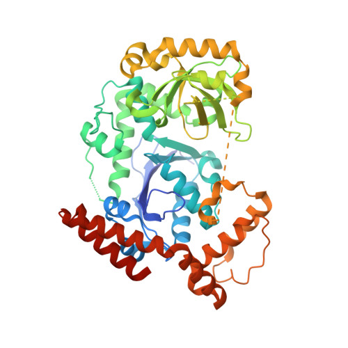
Structure of Escherichia coli exonuclease I in complex with thymidine 5'-monophosphate.

Busam, R.D.

(2008) Acta Crystallogr D Biol Crystallogr 64: 206-210

  • DOI: https://doi.org/10.1107/S090744490706012X
  • Primary Citation of Related Structures:  
    2QXF

  • PubMed Abstract: 

    In Escherichia coli, exonuclease I (ExoI) is a monomeric processive 3'-5' exonuclease that degrades single-stranded DNA. The enzyme has been implicated as primarily being involved in repairing frameshift mutations. The structure of the enzyme has previously been determined in a hexagonal space group at 2.4 A resolution. Here, the structure of ExoI in complex with a nucleotide product, thymidine 5'-monophosphate, is described in an orthorhombic space group at 1.5 A resolution. This new high-resolution structure provides some insight into the interactions involved in binding a nucleotide product. The conserved active site contains a unique metal-binding position when compared with orthologous sites in the Klenow fragment, T4 DNA polymerase and dnaQ. This unique difference is proposed to be a consequence of the repositioning of an important histidine, His181, away from the active site.


  • Organizational Affiliation

    Institute of Molecular Biology, Howard Hughes Medical Institute and Department of Physics, 1229 University of Oregon, Eugene, OR 97403-1229, USA. robert.busam@ki.se


Macromolecules
Find similar proteins by:  (by identity cutoff)  |  3D Structure
Entity ID: 1
MoleculeChains Sequence LengthOrganismDetailsImage
Exodeoxyribonuclease I482Escherichia coli K-12Mutation(s): 0 
Gene Names: sbcBcpeAxonAb2011JW1993
EC: 3.1.11.1
UniProt
Find proteins for P04995 (Escherichia coli (strain K12))
Explore P04995 
Go to UniProtKB:  P04995
Entity Groups  
Sequence Clusters30% Identity50% Identity70% Identity90% Identity95% Identity100% Identity
UniProt GroupP04995
Sequence Annotations
Expand
  • Reference Sequence
Experimental Data & Validation

Experimental Data

  • Method: X-RAY DIFFRACTION
  • Resolution: 1.50 Å
  • R-Value Free: 0.224 
  • R-Value Work: 0.199 
  • R-Value Observed: 0.200 
  • Space Group: P 21 21 21
Unit Cell:
Length ( Å )Angle ( ˚ )
a = 52.63α = 90
b = 91.849β = 90
c = 102.752γ = 90
Software Package:
Software NamePurpose
DENZOdata reduction
SCALEPACKdata scaling
EPMRphasing
REFMACrefinement
PDB_EXTRACTdata extraction
ADSCdata collection
HKL-2000data reduction

Structure Validation

View Full Validation Report


Ligand Structure Quality Assessment 


Entry History 

Deposition Data

  • Released Date: 2008-01-22 
  • Deposition Author(s): Busam, R.D.

Revision History  (Full details and data files)

  • Version 1.0: 2008-01-22
    Type: Initial release
  • Version 1.1: 2011-07-13
    Changes: Advisory, Source and taxonomy, Version format compliance
  • Version 1.2: 2017-10-25
    Changes: Refinement description
  • Version 1.3: 2024-02-21
    Changes: Data collection, Database references, Derived calculations