2QX1

Crystal structure of the complex between mycobacterium tuberculosis beta-ketoacyl-acyl carrier protein synthase III (FABH) and decyl-COA disulfide


Experimental Data Snapshot

  • Method: X-RAY DIFFRACTION
  • Resolution: 2.60 Å
  • R-Value Free: 0.316 
  • R-Value Work: 0.242 
  • R-Value Observed: 0.250 

wwPDB Validation   3D Report Full Report


Ligand Structure Quality Assessment 


This is version 1.3 of the entry. See complete history


Literature

Probing reactivity and substrate specificity of both subunits of the dimeric Mycobacterium tuberculosis FabH using alkyl-CoA disulfide inhibitors and acyl-CoA substrates

Sachdeva, S.Musayev, F.Alhamadsheh, M.M.Neel Scarsdale, J.Tonie Wright, H.Reynolds, K.A.

(2008) Bioorg Chem 36: 85-90

  • DOI: https://doi.org/10.1016/j.bioorg.2007.11.001
  • Primary Citation of Related Structures:  
    2QX1

  • PubMed Abstract: 

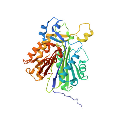
    The dimeric Mycobacterium tuberculosis FabH (mtFabH) catalyses a Claisen-type condensation between an acyl-CoA and malonyl-acyl carrier protein (ACP) to initiate the Type II fatty acid synthase cycle. To analyze the initial covalent acylation of mtFabH with acyl-CoA, we challenged it with mixture of C6-C20 acyl-CoAs and the ESI-MS analysis showed reaction at both subunits and a strict specificity for C12 acyl CoA. Crystallographic and ESI-MS studies of mtFabH with a decyl-CoA disulfide inhibitor revealed a decyl chain bound in acyl-binding channels of both subunits through disulfide linkage to the active site cysteine. These data provide the first unequivocal evidence that both subunits of mtFabH can react with substrates or inhibitor. The discrepancy between the observed C12 acyl-CoA substrate specificity in the initial acylation step and the higher catalytic efficiency of mtFabH for C18-C20 acyl-CoA substrates in the overall mtFabH catalyzed reaction suggests a role for M. tuberculosis ACP as a specificity determinant in this reaction.


  • Organizational Affiliation

    Department of Chemistry, Portland State University, Portland, OR, USA.


Macromolecules
Find similar proteins by:  (by identity cutoff)  |  3D Structure
Entity ID: 1
MoleculeChains Sequence LengthOrganismDetailsImage
Beta-ketoacyl-ACP synthase III
A, B
335Mycobacterium tuberculosisMutation(s): 0 
Gene Names: fabH
EC: 2.3.1.41
UniProt
Find proteins for P9WNG3 (Mycobacterium tuberculosis (strain ATCC 25618 / H37Rv))
Explore P9WNG3 
Go to UniProtKB:  P9WNG3
Entity Groups  
Sequence Clusters30% Identity50% Identity70% Identity90% Identity95% Identity100% Identity
UniProt GroupP9WNG3
Sequence Annotations
Expand
  • Reference Sequence
Experimental Data & Validation

Experimental Data

  • Method: X-RAY DIFFRACTION
  • Resolution: 2.60 Å
  • R-Value Free: 0.316 
  • R-Value Work: 0.242 
  • R-Value Observed: 0.250 
  • Space Group: C 2 2 21
Unit Cell:
Length ( Å )Angle ( ˚ )
a = 67.649α = 90
b = 89.365β = 90
c = 232.006γ = 90
Software Package:
Software NamePurpose
REFMACrefinement
CNSrefinement
CrystalCleardata collection
CrystalCleardata reduction
d*TREKdata scaling
CNSphasing

Structure Validation

View Full Validation Report


Ligand Structure Quality Assessment 


Entry History 

Deposition Data

Revision History  (Full details and data files)

  • Version 1.0: 2008-03-18
    Type: Initial release
  • Version 1.1: 2011-07-13
    Changes: Advisory, Version format compliance
  • Version 1.2: 2019-07-24
    Changes: Data collection, Derived calculations, Refinement description
  • Version 1.3: 2023-08-30
    Changes: Data collection, Database references, Derived calculations, Refinement description