2QRW

Crystal structure of Mycobacterium tuberculosis trHbO WG8F mutant


Experimental Data Snapshot

  • Method: X-RAY DIFFRACTION
  • Resolution: 1.93 Å
  • R-Value Free: 0.215 
  • R-Value Work: 0.176 
  • R-Value Observed: 0.178 

wwPDB Validation   3D Report Full Report


Ligand Structure Quality Assessment 


This is version 1.4 of the entry. See complete history


Literature

The Roles of Tyr(CD1) and Trp(G8) in Mycobacterium tuberculosis Truncated Hemoglobin O in Ligand Binding and on the Heme Distal Site Architecture

Ouellet, H.Milani, M.LaBarre, M.Bolognesi, M.Couture, M.Guertin, M.

(2007) Biochemistry 46: 11440-11450

  • DOI: https://doi.org/10.1021/bi7010288
  • Primary Citation of Related Structures:  
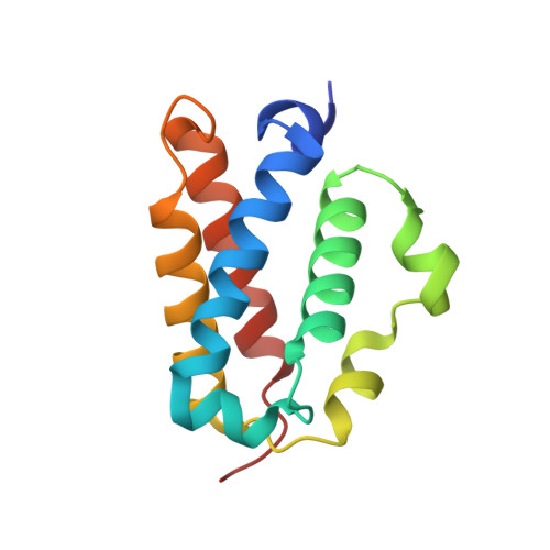
    2QRW

  • PubMed Abstract: 

    The crystal structure of the cyano-met form of Mt-trHbO revealed two unusual distal residues Y(CD1) and W(G8) forming a hydrogen-bond network with the heme-bound ligand [Milani, M., et al. (2003) Proc. Natl. Acad. Sci. U.S.A. 100, 5766-5771]. W(G8) is an invariant residue in group II and group III trHbs and has no counterpart in other globins. A previous study reported that changing Y(CD1) for a Phe causes a significant increase in the O2 combination rate, but almost no change in the O2 dissociation rate [Ouellet, H., et al. (2003) Biochemistry 42, 5764-5774]. Here we investigated the role of the W(G8) in ligand binding by using resonance Raman spectroscopy, stopped-flow spectrophotometry, and X-ray crystallography. For this purpose, W(G8) was changed, by site-directed mutagenesis, to a Phe in both the wild-type protein and the mutant Y(CD1)F to create the single mutant W(G8)F and the double mutant Y(CD1)F/W(G8)F, respectively. Resonance Raman results suggest that W(G8) interacts with the heme-bound O2 and CO, as evidenced by the increase of the Fe-O2 stretching mode from 559 to 564 cm-1 and by the lower frequency of the Fe-CO stretching modes (514 and 497 cm-1) compared to that of the wild-type protein. Mutation of W(G8) to Phe indicates that this residue controls ligand binding, as evidenced by a dramatic increase of the combination rates of both O2 and CO. Also, the rate of O2 dissociation showed a 90-1000-fold increase in the W(G8)F and Y(CD1)F/W(G8)F mutants, that is in sharp contrast with the values obtained for the other distal mutants Y(B10)F and Y(CD1)F [Ouellet, H., et al. (2003) Biochemistry 42, 5764-5774]. Taken together, these data indicate a pivotal role for the W(G8) residue in O2 binding and stabilization.


  • Organizational Affiliation

    Department of Biochemistry and Microbiology, Laval University, Quebec, Canada, G1K 7P4.


Macromolecules
Find similar proteins by:  (by identity cutoff)  |  3D Structure
Entity ID: 1
MoleculeChains Sequence LengthOrganismDetailsImage
Hemoglobin-like protein HbO
A, B, C, D, E
A, B, C, D, E, F, G, H, I, J, K, L
128Mycobacterium tuberculosisMutation(s): 1 
Gene Names: glbO
Membrane Entity: Yes 
UniProt
Find proteins for P9WN23 (Mycobacterium tuberculosis (strain ATCC 25618 / H37Rv))
Explore P9WN23 
Go to UniProtKB:  P9WN23
Entity Groups  
Sequence Clusters30% Identity50% Identity70% Identity90% Identity95% Identity100% Identity
UniProt GroupP9WN23
Sequence Annotations
Expand
  • Reference Sequence
Small Molecules
Ligands 3 Unique
IDChains Name / Formula / InChI Key2D Diagram3D Interactions
HEM
Query on HEM

Download Ideal Coordinates CCD File 
BB [auth K]
CA [auth D]
GA [auth E]
GB [auth L]
LA [auth F]
BB [auth K],
CA [auth D],
GA [auth E],
GB [auth L],
LA [auth F],
PA [auth G],
R [auth A],
SA [auth H],
VA [auth I],
W [auth B],
Y [auth C],
YA [auth J]
PROTOPORPHYRIN IX CONTAINING FE
C34 H32 Fe N4 O4
KABFMIBPWCXCRK-RGGAHWMASA-L
SO4
Query on SO4

Download Ideal Coordinates CCD File 
AA [auth D]
AB [auth K]
BA [auth D]
DB [auth L]
EA [auth E]
AA [auth D],
AB [auth K],
BA [auth D],
DB [auth L],
EA [auth E],
EB [auth L],
FA [auth E],
FB [auth L],
IA [auth F],
JA [auth F],
KA [auth F],
N [auth A],
NA [auth G],
O [auth A],
OA [auth G],
P [auth A],
Q [auth A],
RA [auth H],
T [auth B],
U [auth B],
UA [auth I],
V [auth B],
XA [auth J]
SULFATE ION
O4 S
QAOWNCQODCNURD-UHFFFAOYSA-L
CYN
Query on CYN

Download Ideal Coordinates CCD File 
CB [auth L]
DA [auth E]
HA [auth F]
M [auth A]
MA [auth G]
CB [auth L],
DA [auth E],
HA [auth F],
M [auth A],
MA [auth G],
QA [auth H],
S [auth B],
TA [auth I],
WA [auth J],
X [auth C],
Z [auth D],
ZA [auth K]
CYANIDE ION
C N
XFXPMWWXUTWYJX-UHFFFAOYSA-N
Experimental Data & Validation

Experimental Data

  • Method: X-RAY DIFFRACTION
  • Resolution: 1.93 Å
  • R-Value Free: 0.215 
  • R-Value Work: 0.176 
  • R-Value Observed: 0.178 
  • Space Group: I 41 2 2
Unit Cell:
Length ( Å )Angle ( ˚ )
a = 187.312α = 90
b = 187.312β = 90
c = 274.467γ = 90
Software Package:
Software NamePurpose
REFMACrefinement
ADSCdata collection
MOSFLMdata reduction
SCALAdata scaling
MOLREPphasing

Structure Validation

View Full Validation Report


Ligand Structure Quality Assessment 


Entry History 

Deposition Data

Revision History  (Full details and data files)

  • Version 1.0: 2007-11-06
    Type: Initial release
  • Version 1.1: 2011-07-13
    Changes: Version format compliance
  • Version 1.2: 2016-12-28
    Changes: Structure summary
  • Version 1.3: 2021-10-20
    Changes: Database references, Derived calculations
  • Version 1.4: 2023-08-30
    Changes: Data collection, Refinement description