2QKS

Crystal structure of a Kir3.1-prokaryotic Kir channel chimera


Experimental Data Snapshot

  • Method: X-RAY DIFFRACTION
  • Resolution: 2.20 Å
  • R-Value Free: 0.256 
  • R-Value Work: 0.231 
  • R-Value Observed: 0.232 

wwPDB Validation   3D Report Full Report


Ligand Structure Quality Assessment 


This is version 1.5 of the entry. See complete history


Literature

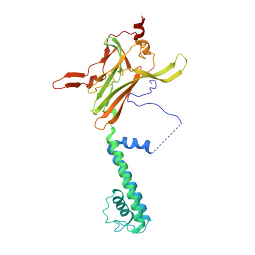
Crystal structure of a Kir3.1-prokaryotic Kir channel chimera.

Nishida, M.Cadene, M.Chait, B.T.Mackinnon, R.

(2007) EMBO J 26: 4005-4015

  • DOI: https://doi.org/10.1038/sj.emboj.7601828
  • Primary Citation of Related Structures:  
    2QKS

  • PubMed Abstract: 

    The Kir3.1 K(+) channel participates in heart rate control and neuronal excitability through G-protein and lipid signaling pathways. Expression in Escherichia coli has been achieved by replacing three fourths of the transmembrane pore with the pore of a prokaryotic Kir channel, leaving the cytoplasmic pore and membrane interfacial regions of Kir3.1 origin. Two structures were determined at 2.2 A. The selectivity filter is identical to the Streptomyces lividans K(+) channel within error of measurement (r.m.s.d.<0.2 A), suggesting that K(+) selectivity requires extreme conservation of three-dimensional structure. Multiple K(+) ions reside within the pore and help to explain voltage-dependent Mg(2+) and polyamine blockade and strong rectification. Two constrictions, at the inner helix bundle and at the apex of the cytoplasmic pore, may function as gates: in one structure the apex is open and in the other, it is closed. Gating of the apex is mediated by rigid-body movements of the cytoplasmic pore subunits. Phosphatidylinositol 4,5-biphosphate-interacting residues suggest a possible mechanism by which the signaling lipid regulates the cytoplasmic pore.


  • Organizational Affiliation

    Laboratory of Molecular Neurobiology and Biophysics, Howard Hughes Medical Institute, Rockefeller University, New York, NY 10065, USA.


Macromolecules
Find similar proteins by:  (by identity cutoff)  |  3D Structure
Entity ID: 1
MoleculeChains Sequence LengthOrganismDetailsImage
Kir3.1-prokaryotic Kir channel chimera
A, B
321Mus musculusParaburkholderia xenovorans LB400Mutation(s): 1 
Membrane Entity: Yes 
UniProt & NIH Common Fund Data Resources
Find proteins for P63250 (Mus musculus)
Explore P63250 
Go to UniProtKB:  P63250
IMPC:  MGI:104742
Find proteins for Q146M9 (Paraburkholderia xenovorans (strain LB400))
Explore Q146M9 
Go to UniProtKB:  Q146M9
Entity Groups  
Sequence Clusters30% Identity50% Identity70% Identity90% Identity95% Identity100% Identity
UniProt GroupsQ146M9P63250
Sequence Annotations
Expand
  • Reference Sequence
Small Molecules
Ligands 2 Unique
IDChains Name / Formula / InChI Key2D Diagram3D Interactions
BNG
Query on BNG

Download Ideal Coordinates CCD File 
C [auth A]nonyl beta-D-glucopyranoside
C15 H30 O6
QFAPUKLCALRPLH-UXXRCYHCSA-N
K
Query on K

Download Ideal Coordinates CCD File 
D [auth A]
E [auth A]
F [auth A]
G [auth A]
H [auth A]
D [auth A],
E [auth A],
F [auth A],
G [auth A],
H [auth A],
I [auth A],
J [auth A],
K [auth B],
L [auth B],
M [auth B],
N [auth B],
O [auth B]
POTASSIUM ION
K
NPYPAHLBTDXSSS-UHFFFAOYSA-N
Experimental Data & Validation

Experimental Data

  • Method: X-RAY DIFFRACTION
  • Resolution: 2.20 Å
  • R-Value Free: 0.256 
  • R-Value Work: 0.231 
  • R-Value Observed: 0.232 
  • Space Group: P 4
Unit Cell:
Length ( Å )Angle ( ˚ )
a = 98.416α = 90
b = 98.416β = 90
c = 92.624γ = 90
Software Package:
Software NamePurpose
CNSrefinement
PDB_EXTRACTdata extraction
HKL-2000data collection
HKL-2000data reduction
HKL-2000data scaling
MOLREPphasing

Structure Validation

View Full Validation Report


Ligand Structure Quality Assessment 


Entry History 

Deposition Data

Revision History  (Full details and data files)

  • Version 1.0: 2007-08-28
    Type: Initial release
  • Version 1.1: 2011-07-13
    Changes: Version format compliance
  • Version 1.2: 2011-11-16
    Changes: Atomic model
  • Version 1.3: 2017-06-07
    Changes: Database references, Other, Source and taxonomy
  • Version 1.4: 2020-07-29
    Type: Remediation
    Reason: Carbohydrate remediation
    Changes: Data collection, Derived calculations, Structure summary
  • Version 1.5: 2023-08-30
    Changes: Data collection, Database references, Refinement description, Structure summary