2QKI

Human C3c in complex with the inhibitor compstatin


Experimental Data Snapshot

  • Method: X-RAY DIFFRACTION
  • Resolution: 2.40 Å
  • R-Value Free: 0.281 
  • R-Value Work: 0.213 
  • R-Value Observed: 0.216 

wwPDB Validation   3D Report Full Report


This is version 2.1 of the entry. See complete history


Literature

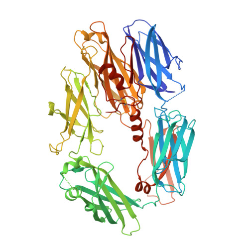
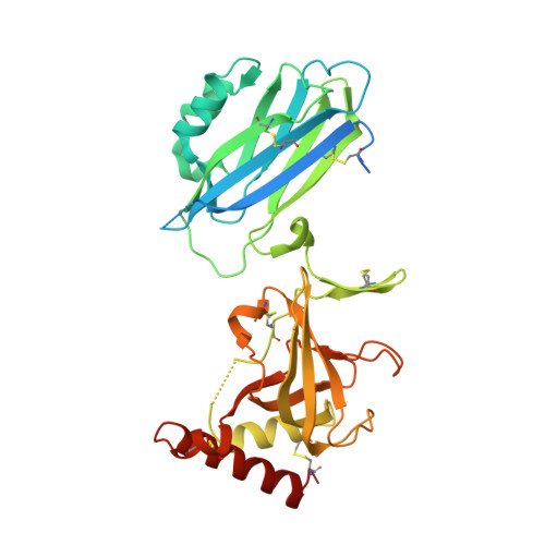
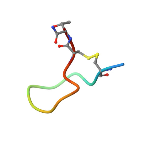
Structure of compstatin in complex with complement component C3c reveals a new mechanism of complement inhibition.

Janssen, B.J.Halff, E.F.Lambris, J.D.Gros, P.

(2007) J Biol Chem 282: 29241-29247

  • DOI: https://doi.org/10.1074/jbc.M704587200
  • Primary Citation of Related Structures:  
    2QKI

  • PubMed Abstract: 

    Undesired complement activation is a major cause of tissue injury in various pathological conditions and contributes to several immune complex diseases. Compstatin, a 13-residue peptide, is an effective inhibitor of the activation of complement component C3 and thus blocks a central and crucial step in the complement cascade. The precise binding site on C3, the structure in the bound form, and the exact mode of action of compstatin are unknown. Here we present the crystal structure of compstatin in complex with C3c, a major proteolytic fragment of C3. The structure reveals that the compstatin-binding site is formed by the macroglobulin (MG) domains 4 and 5. This binding site is part of the structurally stable MG-ring formed by domains MG 1-6 and is far away from any other known binding site on C3. Compstatin does not alter the conformation of C3c, whereas compstatin itself undergoes a large conformational change upon binding. We propose a model in which compstatin sterically hinders the access of the substrate C3 to the convertase complexes, thus blocking complement activation and amplification. These insights are instrumental for further development of compstatin as a potential therapeutic.


  • Organizational Affiliation

    Crystal and Structural Chemistry, Bijvoet Center for Biomolecular Research, Department of Chemistry, Faculty of Sciences, Utrecht University, 3584 CH Utrecht, The Netherlands.


Macromolecules
Find similar proteins by:  (by identity cutoff)  |  3D Structure
Entity ID: 1
MoleculeChains Sequence LengthOrganismDetailsImage
Complement C3
A, D
643Homo sapiensMutation(s): 0 
UniProt & NIH Common Fund Data Resources
Find proteins for P01024 (Homo sapiens)
Explore P01024 
Go to UniProtKB:  P01024
PHAROS:  P01024
GTEx:  ENSG00000125730 
Entity Groups  
Sequence Clusters30% Identity50% Identity70% Identity90% Identity95% Identity100% Identity
UniProt GroupP01024
Sequence Annotations
Expand
  • Reference Sequence
Find similar proteins by:  (by identity cutoff)  |  3D Structure
Entity ID: 2
MoleculeChains Sequence LengthOrganismDetailsImage
Complement C3
B, E
188Homo sapiensMutation(s): 0 
UniProt & NIH Common Fund Data Resources
Find proteins for P01024 (Homo sapiens)
Explore P01024 
Go to UniProtKB:  P01024
PHAROS:  P01024
GTEx:  ENSG00000125730 
Entity Groups  
Sequence Clusters30% Identity50% Identity70% Identity90% Identity95% Identity100% Identity
UniProt GroupP01024
Sequence Annotations
Expand
  • Reference Sequence
Find similar proteins by:  (by identity cutoff)  |  3D Structure
Entity ID: 3
MoleculeChains Sequence LengthOrganismDetailsImage
Complement C3
C, F
343Homo sapiensMutation(s): 0 
UniProt & NIH Common Fund Data Resources
Find proteins for P01024 (Homo sapiens)
Explore P01024 
Go to UniProtKB:  P01024
PHAROS:  P01024
GTEx:  ENSG00000125730 
Entity Groups  
Sequence Clusters30% Identity50% Identity70% Identity90% Identity95% Identity100% Identity
UniProt GroupP01024
Sequence Annotations
Expand
  • Reference Sequence

Find similar proteins by:  Sequence   |   3D Structure  

Entity ID: 4
MoleculeChains Sequence LengthOrganismDetailsImage
compstatin
G, H
15N/AMutation(s): 0 
Entity Groups  
Sequence Clusters30% Identity50% Identity70% Identity90% Identity95% Identity100% Identity
Sequence Annotations
Expand
  • Reference Sequence
Oligosaccharides

Help

Entity ID: 5
MoleculeChains Length2D Diagram Glycosylation3D Interactions
2-acetamido-2-deoxy-beta-D-glucopyranose-(1-4)-2-acetamido-2-deoxy-beta-D-glucopyranose
I, J
2N-Glycosylation
Glycosylation Resources
GlyTouCan:  G42666HT
GlyCosmos:  G42666HT
GlyGen:  G42666HT
Small Molecules
Ligands 3 Unique
IDChains Name / Formula / InChI Key2D Diagram3D Interactions
GOL
Query on GOL

Download Ideal Coordinates CCD File 
AB [auth D]
BA [auth B]
BB [auth D]
CB [auth D]
EA [auth C]
AB [auth D],
BA [auth B],
BB [auth D],
CB [auth D],
EA [auth C],
GB [auth E],
HB [auth E],
LB [auth F],
MB [auth H],
T [auth A],
U [auth A],
V [auth A],
W [auth A],
WA [auth D],
X [auth A],
XA [auth D],
YA [auth D],
ZA [auth D]
GLYCEROL
C3 H8 O3
PEDCQBHIVMGVHV-UHFFFAOYSA-N
BR
Query on BR

Download Ideal Coordinates CCD File 
AA [auth B]
DA [auth C]
DB [auth E]
EB [auth E]
FB [auth E]
AA [auth B],
DA [auth C],
DB [auth E],
EB [auth E],
FB [auth E],
HA [auth D],
IA [auth D],
JA [auth D],
JB [auth F],
KA [auth D],
KB [auth F],
L [auth A],
LA [auth D],
M [auth A],
MA [auth D],
N [auth A],
NA [auth D],
O [auth A],
OA [auth D],
P [auth A],
PA [auth D],
Q [auth A],
QA [auth D],
R [auth A],
RA [auth D],
S [auth A],
SA [auth D],
TA [auth D],
UA [auth D],
VA [auth D],
Y [auth B],
Z [auth B]
BROMIDE ION
Br
CPELXLSAUQHCOX-UHFFFAOYSA-M
K
Query on K

Download Ideal Coordinates CCD File 
CA [auth C],
FA [auth D],
GA [auth D],
IB [auth F],
K [auth A]
POTASSIUM ION
K
NPYPAHLBTDXSSS-UHFFFAOYSA-N
Experimental Data & Validation

Experimental Data

  • Method: X-RAY DIFFRACTION
  • Resolution: 2.40 Å
  • R-Value Free: 0.281 
  • R-Value Work: 0.213 
  • R-Value Observed: 0.216 
  • Space Group: P 1 21 1
Unit Cell:
Length ( Å )Angle ( ˚ )
a = 85.809α = 90
b = 124.754β = 95.08
c = 127.367γ = 90
Software Package:
Software NamePurpose
REFMACrefinement
DNAdata collection
MOSFLMdata reduction
SCALAdata scaling
PHASERphasing

Structure Validation

View Full Validation Report


Entry History 

Deposition Data

Revision History  (Full details and data files)

  • Version 1.0: 2007-08-14
    Type: Initial release
  • Version 1.1: 2011-07-13
    Changes: Atomic model, Database references, Derived calculations, Non-polymer description, Structure summary, Version format compliance
  • Version 2.0: 2020-07-29
    Type: Remediation
    Reason: Carbohydrate remediation
    Changes: Advisory, Atomic model, Data collection, Derived calculations, Structure summary
  • Version 2.1: 2023-08-30
    Changes: Data collection, Database references, Refinement description, Structure summary