2QH7

MitoNEET is a uniquely folded 2Fe-2S outer mitochondrial membrane protein stabilized by pioglitazone


Experimental Data Snapshot

  • Method: X-RAY DIFFRACTION
  • Resolution: 1.50 Å
  • R-Value Free: 0.223 
  • R-Value Work: 0.182 
  • R-Value Observed: 0.184 

wwPDB Validation   3D Report Full Report


This is version 1.3 of the entry. See complete history


Literature

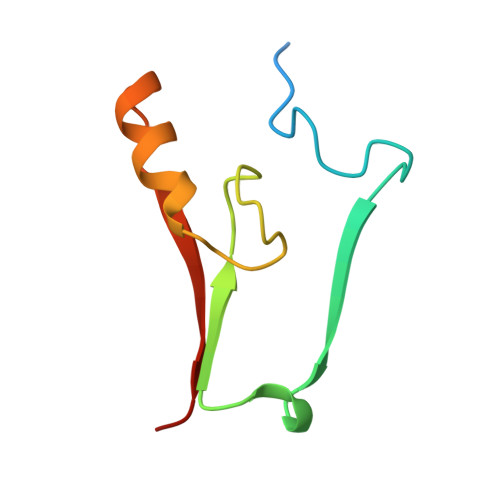
MitoNEET is a uniquely folded 2Fe 2S outer mitochondrial membrane protein stabilized by pioglitazone.

Paddock, M.L.Wiley, S.E.Axelrod, H.L.Cohen, A.E.Roy, M.Abresch, E.C.Capraro, D.Murphy, A.N.Nechushtai, R.Dixon, J.E.Jennings, P.A.

(2007) Proc Natl Acad Sci U S A 104: 14342-14347

  • DOI: https://doi.org/10.1073/pnas.0707189104
  • Primary Citation of Related Structures:  
    2QH7

  • PubMed Abstract: 

    Iron-sulfur (Fe-S) proteins are key players in vital processes involving energy homeostasis and metabolism from the simplest to most complex organisms. We report a 1.5 A x-ray crystal structure of the first identified outer mitochondrial membrane Fe-S protein, mitoNEET. Two protomers intertwine to form a unique dimeric structure that constitutes a new fold to not only the approximately 650 reported Fe-S protein structures but also to all known proteins. We name this motif the NEET fold. The protomers form a two-domain structure: a beta-cap domain and a cluster-binding domain that coordinates two acid-labile 2Fe-2S clusters. Binding of pioglitazone, an insulin-sensitizing thiazolidinedione used in the treatment of type 2 diabetes, stabilizes the protein against 2Fe-2S cluster release. The biophysical properties of mitoNEET suggest that it may participate in a redox-sensitive signaling and/or in Fe-S cluster transfer.


  • Organizational Affiliation

    Department of Physics, University of California at San Diego, La Jolla, CA 92093, USA.


Macromolecules
Find similar proteins by:  (by identity cutoff)  |  3D Structure
Entity ID: 1
MoleculeChains Sequence LengthOrganismDetailsImage
Zinc finger CDGSH-type domain 1
A, B
76Homo sapiensMutation(s): 0 
Gene Names: ZCD1
UniProt & NIH Common Fund Data Resources
Find proteins for Q9NZ45 (Homo sapiens)
Explore Q9NZ45 
Go to UniProtKB:  Q9NZ45
PHAROS:  Q9NZ45
GTEx:  ENSG00000122873 
Entity Groups  
Sequence Clusters30% Identity50% Identity70% Identity90% Identity95% Identity100% Identity
UniProt GroupQ9NZ45
Sequence Annotations
Expand
  • Reference Sequence
Small Molecules
Experimental Data & Validation

Experimental Data

  • Method: X-RAY DIFFRACTION
  • Resolution: 1.50 Å
  • R-Value Free: 0.223 
  • R-Value Work: 0.182 
  • R-Value Observed: 0.184 
  • Space Group: P 21 21 21
Unit Cell:
Length ( Å )Angle ( ˚ )
a = 46.806α = 90
b = 49.621β = 90
c = 59.006γ = 90
Software Package:
Software NamePurpose
XSCALEdata scaling
SOLVEphasing
RESOLVEphasing
REFMACrefinement
PDB_EXTRACTdata extraction
Blu-Icedata collection
MOSFLMdata reduction
XDSdata reduction
SCALAdata scaling

Structure Validation

View Full Validation Report


Entry History 

Revision History  (Full details and data files)

  • Version 1.0: 2007-08-21
    Type: Initial release
  • Version 1.1: 2011-07-13
    Changes: Advisory, Derived calculations, Version format compliance
  • Version 1.2: 2018-01-24
    Changes: Structure summary
  • Version 1.3: 2024-02-21
    Changes: Data collection, Database references, Derived calculations, Refinement description