2Q8A

Structure of the malaria antigen AMA1 in complex with a growth-inhibitory antibody


Experimental Data Snapshot

  • Method: X-RAY DIFFRACTION
  • Resolution: 2.40 Å
  • R-Value Free: 0.244 
  • R-Value Work: 0.202 
  • R-Value Observed: 0.204 

wwPDB Validation   3D Report Full Report


This is version 1.3 of the entry. See complete history


Literature

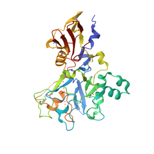
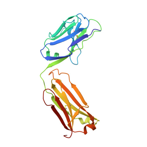
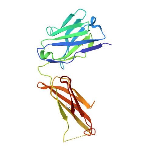
Structure of the malaria antigen AMA1 in complex with a growth-inhibitory antibody.

Coley, A.M.Gupta, A.Murphy, V.J.Bai, T.Kim, H.Foley, M.Anders, R.F.Batchelor, A.H.

(2007) PLoS Pathog 3: 1308-1319

  • DOI: https://doi.org/10.1371/journal.ppat.0030138
  • Primary Citation of Related Structures:  
    2Q8A, 2Q8B

  • PubMed Abstract: 

    Identifying functionally critical regions of the malaria antigen AMA1 (apical membrane antigen 1) is necessary to understand the significance of the polymorphisms within this antigen for vaccine development. The crystal structure of AMA1 in complex with the Fab fragment of inhibitory monoclonal antibody 1F9 reveals that 1F9 binds to the AMA1 solvent-exposed hydrophobic trough, confirming its importance. 1F9 uses the heavy and light chain complementarity-determining regions (CDRs) to wrap around the polymorphic loops adjacent to the trough, but uses a ridge of framework residues to bind to the hydrophobic trough. The resulting 1F9-AMA1-combined buried surface of 2,470 A(2) is considerably larger than previously reported Fab-antigen interfaces. Mutations of polymorphic AMA1 residues within the 1F9 epitope disrupt 1F9 binding and dramatically reduce the binding of affinity-purified human antibodies. Moreover, 1F9 binding to AMA1 is competed by naturally acquired human antibodies, confirming that the 1F9 epitope is a frequent target of immunological attack.


  • Organizational Affiliation

    Cooperative Research Center for Diagnostics, Department of Biochemistry, La Trobe University, Victoria, Australia.


Macromolecules
Find similar proteins by:  (by identity cutoff)  |  3D Structure
Entity ID: 1
MoleculeChains Sequence LengthOrganismDetailsImage
Apical membrane antigen 1336Plasmodium falciparum 3D7Mutation(s): 0 
Gene Names: AMA1
UniProt
Find proteins for Q7KQK5 (Plasmodium falciparum (isolate 3D7))
Explore Q7KQK5 
Go to UniProtKB:  Q7KQK5
Entity Groups  
Sequence Clusters30% Identity50% Identity70% Identity90% Identity95% Identity100% Identity
UniProt GroupQ7KQK5
Sequence Annotations
Expand
  • Reference Sequence
Find similar proteins by:  (by identity cutoff)  |  3D Structure
Entity ID: 2
MoleculeChains Sequence LengthOrganismDetailsImage
1F9 light chainB [auth L]214Mus musculusMutation(s): 0 
Entity Groups  
Sequence Clusters30% Identity50% Identity70% Identity90% Identity95% Identity100% Identity
Sequence Annotations
Expand
  • Reference Sequence
Find similar proteins by:  (by identity cutoff)  |  3D Structure
Entity ID: 3
MoleculeChains Sequence LengthOrganismDetailsImage
1F9 heavy chainC [auth H]210Mus musculusMutation(s): 0 
Entity Groups  
Sequence Clusters30% Identity50% Identity70% Identity90% Identity95% Identity100% Identity
Sequence Annotations
Expand
  • Reference Sequence
Experimental Data & Validation

Experimental Data

  • Method: X-RAY DIFFRACTION
  • Resolution: 2.40 Å
  • R-Value Free: 0.244 
  • R-Value Work: 0.202 
  • R-Value Observed: 0.204 
  • Space Group: C 1 2 1
Unit Cell:
Length ( Å )Angle ( ˚ )
a = 189.762α = 90
b = 51.203β = 111.09
c = 91.518γ = 90
Software Package:
Software NamePurpose
REFMACrefinement
CrystalCleardata collection
d*TREKdata reduction
d*TREKdata scaling
PHASERphasing

Structure Validation

View Full Validation Report


Entry History 

Deposition Data

Revision History  (Full details and data files)

  • Version 1.0: 2007-10-09
    Type: Initial release
  • Version 1.1: 2011-07-13
    Changes: Advisory, Source and taxonomy, Version format compliance
  • Version 1.2: 2017-12-20
    Changes: Database references
  • Version 1.3: 2023-08-30
    Changes: Data collection, Database references, Refinement description