2Q32

Crystal structure of human heme oxygenase-2 C127A (HO-2)


Experimental Data Snapshot

  • Method: X-RAY DIFFRACTION
  • Resolution: 2.40 Å
  • R-Value Free: 0.253 
  • R-Value Work: 0.201 
  • R-Value Observed: 0.204 

wwPDB Validation   3D Report Full Report


Ligand Structure Quality Assessment 


This is version 1.5 of the entry. See complete history


Literature

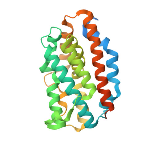
Comparison of Apo- and Heme-bound Crystal Structures of a Truncated Human Heme Oxygenase-2.

Bianchetti, C.M.Yi, L.Ragsdale, S.W.Phillips Jr., G.N.

(2007) J Biol Chem 282: 37624-37631

  • DOI: https://doi.org/10.1074/jbc.M707396200
  • Primary Citation of Related Structures:  
    2Q32, 2QPP, 2RGZ

  • PubMed Abstract: 

    Heme oxygenase (HO) catalyzes the first step in the heme degradation pathway. The crystal structures of apo- and heme-bound truncated human HO-2 reveal a primarily alpha-helical architecture similar to that of human HO-1 and other known HOs. Proper orientation of heme in HO-2 is required for the regioselective oxidation of the alpha-mesocarbon. This is accomplished by interactions within the heme binding pocket, which is made up of two helices. The iron coordinating residue, His(45), resides on the proximal helix. The distal helix contains highly conserved glycine residues that allow the helix to flex and interact with the bound heme. Tyr(154), Lys(199), and Arg(203) orient the heme through direct interactions with the heme propionates. The rearrangements of side chains in heme-bound HO-2 compared with apoHO-2 further elucidate HO-2 heme interactions.


  • Organizational Affiliation

    Graduate Program in Biophysics, Center for Eukaryotic Structural Genomics, University of Wisconsin, Madison, WI 53706, USA.


Macromolecules
Find similar proteins by:  (by identity cutoff)  |  3D Structure
Entity ID: 1
MoleculeChains Sequence LengthOrganismDetailsImage
Heme oxygenase 2
A, B
264Homo sapiensMutation(s): 1 
Gene Names: Hs.284279HMOX2HO2
EC: 1.14.99.3
UniProt & NIH Common Fund Data Resources
Find proteins for P30519 (Homo sapiens)
Explore P30519 
Go to UniProtKB:  P30519
PHAROS:  P30519
GTEx:  ENSG00000103415 
Entity Groups  
Sequence Clusters30% Identity50% Identity70% Identity90% Identity95% Identity100% Identity
UniProt GroupP30519
Sequence Annotations
Expand
  • Reference Sequence
Experimental Data & Validation

Experimental Data

  • Method: X-RAY DIFFRACTION
  • Resolution: 2.40 Å
  • R-Value Free: 0.253 
  • R-Value Work: 0.201 
  • R-Value Observed: 0.204 
  • Space Group: P 21 21 21
Unit Cell:
Length ( Å )Angle ( ˚ )
a = 75.769α = 90
b = 86.017β = 90
c = 97.753γ = 90
Software Package:
Software NamePurpose
DENZOdata reduction
SCALEPACKdata scaling
MOLREPphasing
REFMACrefinement
PDB_EXTRACTdata extraction
MAR345data collection
HKL-2000data reduction
HKL-2000data scaling

Structure Validation

View Full Validation Report


Ligand Structure Quality Assessment 


Entry History 

Revision History  (Full details and data files)

  • Version 1.0: 2007-06-05
    Type: Initial release
  • Version 1.1: 2008-01-15
    Changes: Version format compliance
  • Version 1.2: 2011-07-13
    Changes: Advisory, Version format compliance
  • Version 1.3: 2017-10-18
    Changes: Refinement description
  • Version 1.4: 2021-10-20
    Changes: Database references, Derived calculations
  • Version 1.5: 2023-08-30
    Changes: Data collection, Refinement description