Crystal Structure of the Bacterial Ribosomal Decoding Site Complexed with a Synthetic Doubly Functionalized Paromomycin Derivative: a New Specific Binding Mode to an A-Minor Motif Enhances in vitro Antibacterial Activity
Kondo, J., Pachamuthu, K., Francois, B., Szychowski, J., Hanessian, S., Westhof, E.(2007) ChemMedChem 2: 1631-1638
- PubMed: 17722211
- DOI: https://doi.org/10.1002/cmdc.200700113
- Primary Citation of Related Structures:
2PWT - PubMed Abstract:
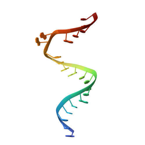
The crystal structure of the complex between oligonucleotide containing the bacterial ribosomal decoding site (A site) and the synthetic paromomycin analogue 1, which contains the gamma-amino-alpha-hydroxybutyryl (L-haba) group at position N1 of ring II (2-DOS ring), and an ether chain with an O-phenethylaminoethyl group at position C2'' of ring III, is reported. Interestingly, next to the paromomycin analogue 1 specifically bound to the A site, a second molecule of 1 with a different conformation is observed at the crystal packing interface which mimics the A-minor interaction between two bulged-out adenines from the A site and the codon-anticodon stem of the mRNA-tRNA complex. Improved antibacterial activity supports the conclusion that analogue 1 might affect protein synthesis on the ribosome in two different ways: 1) specific binding to the A site forces maintenance of the "on" state with two bulged out adenines, and 2) a new binding mode of 1 to an A-minor motif which stabilizes complex formation between the ribosome and the mRNA-tRNA complex regardless of whether the codon-anticodon stem is of the cognate or near-cognate type.
Organizational Affiliation:
Architecture et Réactivité de l'ARN, Université Louis Pasteur, Institut de Biologie Moléculaire et Cellulaire, CNRS, 15 rue René Descartes, 67084 Strasbourg, France.