


Crystal structure of the complex of proteinase K with coumarin at 1.9A resolution
Singh, A.K., Singh, N., Sinha, M., Sharma, S., Kaur, P., Singh, T.P.To be published.
Experimental Data Snapshot
wwPDB Validation 3D Report Full Report

Entity ID: 1 | |||||
|---|---|---|---|---|---|
| Molecule | Chains | Sequence Length | Organism | Details | Image |
| Proteinase K | 279 | Parengyodontium album | Mutation(s): 0 EC: 3.4.21.64 | 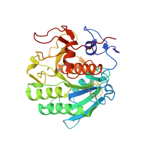 | |
UniProt | |||||
Find proteins for P06873 (Parengyodontium album) Explore P06873 Go to UniProtKB: P06873 | |||||
Entity Groups | |||||
| Sequence Clusters | 30% Identity50% Identity70% Identity90% Identity95% Identity100% Identity | ||||
| UniProt Group | P06873 | ||||
Sequence AnnotationsExpand | |||||
| |||||
| Ligands 3 Unique | |||||
|---|---|---|---|---|---|
| ID | Chains | Name / Formula / InChI Key | 2D Diagram | 3D Interactions | |
| COU Query on COU | E [auth A] | COUMARIN C9 H6 O2 ZYGHJZDHTFUPRJ-UHFFFAOYSA-N |  | ||
| NO3 Query on NO3 | D [auth A] | NITRATE ION N O3 NHNBFGGVMKEFGY-UHFFFAOYSA-N |  | ||
| CA Query on CA | B [auth A], C [auth A] | CALCIUM ION Ca BHPQYMZQTOCNFJ-UHFFFAOYSA-N |  | ||
| Length ( Å ) | Angle ( ˚ ) |
|---|---|
| a = 68.3 | α = 90 |
| b = 68.3 | β = 90 |
| c = 108.384 | γ = 90 |
| Software Name | Purpose |
|---|---|
| REFMAC | refinement |
| MAR345dtb | data collection |
| DENZO | data reduction |
| SCALEPACK | data scaling |
| AMoRE | phasing |
RCSB PDB (citation) is hosted by
RCSB PDB is a member of the

