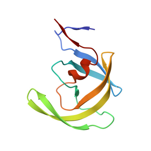
2PSV

Crystal Structure of wild type HIV-1 protease in complex with CARB-KB45


Experimental Data Snapshot

  • Method: X-RAY DIFFRACTION
  • Resolution: 1.75 Å
  • R-Value Free: 0.208 
  • R-Value Work: 0.165 
  • R-Value Observed: 0.167 

wwPDB Validation   3D Report Full Report


Ligand Structure Quality Assessment 


This is version 1.5 of the entry. See complete history


Literature

Design of Mutation-resistant HIV Protease Inhibitors with the Substrate Envelope Hypothesis.

Chellappan, S.Kiran Kumar Reddy, G.S.Ali, A.Nalam, M.N.Anjum, S.G.Cao, H.Kairys, V.Fernandes, M.X.Altman, M.D.Tidor, B.Rana, T.M.Schiffer, C.A.Gilson, M.K.

(2007) Chem Biol Drug Des 69: 298-313

  • DOI: https://doi.org/10.1111/j.1747-0285.2007.00514.x
  • Primary Citation of Related Structures:  
    2PSU, 2PSV

  • PubMed Abstract: 

    There is a clinical need for HIV protease inhibitors that can evade resistance mutations. One possible approach to designing such inhibitors relies upon the crystallographic observation that the substrates of HIV protease occupy a rather constant region within the binding site. In particular, it has been hypothesized that inhibitors which lie within this region will tend to resist clinically relevant mutations. The present study offers the first prospective evaluation of this hypothesis, via computational design of inhibitors predicted to conform to the substrate envelope, followed by synthesis and evaluation against wild-type and mutant proteases, as well as structural studies of complexes of the designed inhibitors with HIV protease. The results support the utility of the substrate envelope hypothesis as a guide to the design of robust protease inhibitors.


  • Organizational Affiliation

    Center for Advanced Research in Biotechnology, University of Maryland, Biotechnology Institute, Rockville, MD 20850, USA.


Macromolecules
Find similar proteins by:  (by identity cutoff)  |  3D Structure
Entity ID: 1
MoleculeChains Sequence LengthOrganismDetailsImage
Protease
A, B
99Human immunodeficiency virus 1Mutation(s): 1 
Gene Names: pol
UniProt
Find proteins for O38707 (Human immunodeficiency virus 1)
Explore O38707 
Go to UniProtKB:  O38707
Entity Groups  
Sequence Clusters30% Identity50% Identity70% Identity90% Identity95% Identity100% Identity
UniProt GroupO38707
Sequence Annotations
Expand
  • Reference Sequence
Small Molecules
Binding Affinity Annotations 
IDSourceBinding Affinity
MUV BindingDB:  2PSV Ki: 58 (nM) from 1 assay(s)
Binding MOAD:  2PSV Ki: 58 (nM) from 1 assay(s)
PDBBind:  2PSV Ki: 58 (nM) from 1 assay(s)
Experimental Data & Validation

Experimental Data

  • Method: X-RAY DIFFRACTION
  • Resolution: 1.75 Å
  • R-Value Free: 0.208 
  • R-Value Work: 0.165 
  • R-Value Observed: 0.167 
  • Space Group: P 21 21 21
Unit Cell:
Length ( Å )Angle ( ˚ )
a = 50.692α = 90
b = 57.593β = 90
c = 61.836γ = 90
Software Package:
Software NamePurpose
REFMACrefinement
HKL-2000data reduction
SCALEPACKdata scaling
AMoREphasing

Structure Validation

View Full Validation Report


Ligand Structure Quality Assessment 


Entry History 

Deposition Data

Revision History  (Full details and data files)

  • Version 1.0: 2007-06-05
    Type: Initial release
  • Version 1.1: 2008-05-01
    Changes: Version format compliance
  • Version 1.2: 2011-07-13
    Changes: Advisory, Refinement description, Version format compliance
  • Version 1.3: 2017-10-18
    Changes: Advisory, Refinement description
  • Version 1.4: 2021-10-20
    Changes: Advisory, Database references, Derived calculations
  • Version 1.5: 2023-08-30
    Changes: Data collection, Refinement description