Synchrotron X-ray data collection and restrained least-squares refinement of the crystal structure of proteinase K at 1.5 A resolution.
Betzel, C., Pal, G.P., Saenger, W.(1988) Acta Crystallogr B 44: 163-172
- PubMed: 3271105
- DOI: https://doi.org/10.1107/s010876818700939x
- Primary Citation of Related Structures:
2PRK - PubMed Abstract:
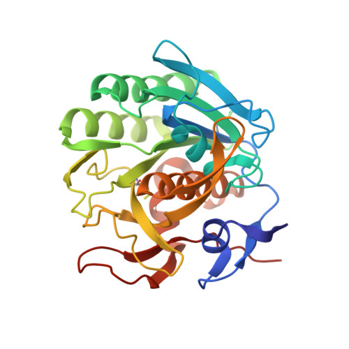
The structure of the serine endopeptidase proteinase K (279 amino acid residues; 28,790 daltons) has been refined by restrained least-squares methods to a conventional R value of 16.7% employing synchrotron film data of 30,812 reflections greater than 3 sigma in the 5.0 to 1.5 A resolution range. During refinement, the molecular structure was restrained to known stereochemistry, with root-mean-square (r.m.s.) deviation of 0.015 A from ideal bond lengths. The average atomic temperature factor, B, is 11.1 A2 for all atoms. The final model comprises 2020 protein atoms and 174 solvent molecules (which were given unit occupancies). Four corrections to the amino acid sequence were made, which were confirmed later by sequence analysis of the proteinase K gene: a deletion of one glycine in position 80; a change of sequence in position 207-208 and insertions of the dipeptide 210-211 and of residue 270. The r.m.s. deviation in the alpha-C atomic positions between the final refined model and the initial model built on the basis of a 3.3 A mini-map is 1.72 A for 227 out of 266 residues, which were originally traced in the mini-map without sequence information. The positions of the remaining 39 residues deviate by more than 8 A from the original ones and are located in regions where extensive revision of the structural model was necessary.
Organizational Affiliation:
Institut für Kristallographie, Freie Universität Berlin, Federal Republic of Germany.