2PKX

E.coli response regulator PhoP receiver domain


Experimental Data Snapshot

  • Method: X-RAY DIFFRACTION
  • Resolution: 2.54 Å
  • R-Value Free: 0.253 
  • R-Value Work: 0.206 
  • R-Value Observed: 0.210 

wwPDB Validation   3D Report Full Report


This is version 1.4 of the entry. See complete history


Literature

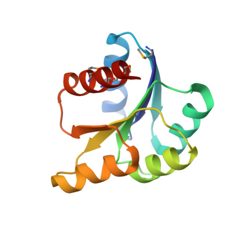
Crystal Structures of the Receiver Domain of the Response Regulator PhoP from Escherichia coli in the Absence and Presence of the Phosphoryl Analog Beryllofluoride.

Bachhawat, P.Stock, A.M.

(2007) J Bacteriol 189: 5987-5995

  • DOI: https://doi.org/10.1128/JB.00049-07
  • Primary Citation of Related Structures:  
    2PKX, 2PL1

  • PubMed Abstract: 

    The response regulator PhoP is part of the PhoQ/PhoP two-component system involved in responses to depletion of extracellular Mg(2+). Here, we report the crystal structures of the receiver domain of Escherichia coli PhoP determined in the absence and presence of the phosphoryl analog beryllofluoride. In the presence of beryllofluoride, the active receiver domain forms a twofold symmetric dimer similar to that seen in structures of other regulatory domains from the OmpR/PhoB family, providing further evidence that members of this family utilize a common mode of dimerization in the active state. In the absence of activating agents, the PhoP receiver domain crystallizes with a similar structure, consistent with the previous observation that high concentrations can promote an active state of PhoP independent of phosphorylation.


  • Organizational Affiliation

    Department of Biochemistry, Center for Advanced Biotechnology and Medicine, 679 Hoes Lane, Piscataway, NJ 08854-5627, USA.


Macromolecules
Find similar proteins by:  (by identity cutoff)  |  3D Structure
Entity ID: 1
MoleculeChains Sequence LengthOrganismDetailsImage
Transcriptional regulatory protein phoP
A, B
121Escherichia coliMutation(s): 5 
Gene Names: phoP
UniProt
Find proteins for P23836 (Escherichia coli (strain K12))
Explore P23836 
Go to UniProtKB:  P23836
Entity Groups  
Sequence Clusters30% Identity50% Identity70% Identity90% Identity95% Identity100% Identity
UniProt GroupP23836
Sequence Annotations
Expand
  • Reference Sequence
Small Molecules
Modified Residues  1 Unique
IDChains TypeFormula2D DiagramParent
MSE
Query on MSE
A, B
L-PEPTIDE LINKINGC5 H11 N O2 SeMET
Experimental Data & Validation

Experimental Data

  • Method: X-RAY DIFFRACTION
  • Resolution: 2.54 Å
  • R-Value Free: 0.253 
  • R-Value Work: 0.206 
  • R-Value Observed: 0.210 
  • Space Group: P 65
Unit Cell:
Length ( Å )Angle ( ˚ )
a = 102.645α = 90
b = 102.645β = 90
c = 62.641γ = 120
Software Package:
Software NamePurpose
REFMACrefinement
ADSCdata collection
DENZOdata reduction
SCALEPACKdata scaling
PHASERphasing

Structure Validation

View Full Validation Report


Entry History 

Deposition Data

  • Released Date: 2007-05-22 
  • Deposition Author(s): Bachhawat, P.
  • This entry supersedes: 2EU6

Revision History  (Full details and data files)

  • Version 1.0: 2007-05-22
    Type: Initial release
  • Version 1.1: 2008-05-01
    Changes: Version format compliance
  • Version 1.2: 2011-07-13
    Changes: Version format compliance
  • Version 1.3: 2021-10-20
    Changes: Database references, Derived calculations
  • Version 1.4: 2024-04-03
    Changes: Data collection, Refinement description