Structures of substrate- and inhibitor-bound adenosine deaminase from a human malaria parasite show a dramatic conformational change and shed light on drug selectivity.
Larson, E.T., Deng, W., Krumm, B.E., Napuli, A., Mueller, N., Van Voorhis, W.C., Buckner, F.S., Fan, E., Lauricella, A., DeTitta, G., Luft, J., Zucker, F., Hol, W.G., Verlinde, C.L., Merritt, E.A.(2008) J Mol Biol 381: 975-988
- PubMed: 18602399
- DOI: https://doi.org/10.1016/j.jmb.2008.06.048
- Primary Citation of Related Structures:
2PGF, 2PGR, 2QVN - PubMed Abstract:
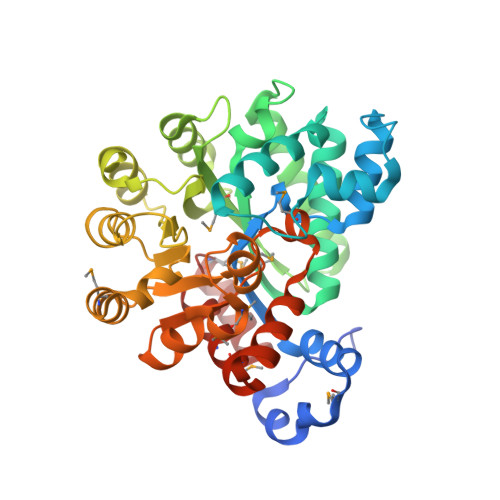
Plasmodium and other apicomplexan parasites are deficient in purine biosynthesis, relying instead on the salvage of purines from their host environment. Therefore, interference with the purine salvage pathway is an attractive therapeutic target. The plasmodial enzyme adenosine deaminase (ADA) plays a central role in purine salvage and, unlike mammalian ADA homologs, has a further secondary role in methylthiopurine recycling. For this reason, plasmodial ADA accepts a wider range of substrates, as it is responsible for deamination of both adenosine and 5'-methylthioadenosine. The latter substrate is not accepted by mammalian ADA homologs. The structural basis for this natural difference in specificity between plasmodial and mammalian ADA has not been well understood. We now report crystal structures of Plasmodium vivax ADA in complex with adenosine, guanosine, and the picomolar inhibitor 2'-deoxycoformycin. These structures highlight a drastic conformational change in plasmodial ADA upon substrate binding that has not been observed for mammalian ADA enzymes. Further, these complexes illuminate the structural basis for the differential substrate specificity and potential drug selectivity between mammalian and parasite enzymes.
Organizational Affiliation:
Medical Structural Genomics of Pathogenic Protozoa Consortium, University of Washington, Seattle, WA 98195-7742, USA.