


Crystal structure of an isomerase from Streptomyces coelicolor
Agarwal, R., Burley, S.K., Swaminathan, S.To be published.
Experimental Data Snapshot
wwPDB Validation 3D Report Full Report

Entity ID: 1 | |||||
|---|---|---|---|---|---|
| Molecule | Chains | Sequence Length | Organism | Details | Image |
| Putative isomerase | 385 | Streptomyces coelicolor A3(2) | Mutation(s): 10 Gene Names: SCO7570, SC5F1.24 | 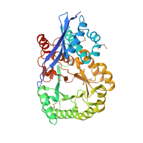 | |
UniProt | |||||
Find proteins for Q9F3A5 (Streptomyces coelicolor (strain ATCC BAA-471 / A3(2) / M145)) Explore Q9F3A5 Go to UniProtKB: Q9F3A5 | |||||
Entity Groups | |||||
| Sequence Clusters | 30% Identity50% Identity70% Identity90% Identity95% Identity100% Identity | ||||
| UniProt Group | Q9F3A5 | ||||
Sequence AnnotationsExpand | |||||
| |||||
| Ligands 1 Unique | |||||
|---|---|---|---|---|---|
| ID | Chains | Name / Formula / InChI Key | 2D Diagram | 3D Interactions | |
| SO4 Query on SO4 | E [auth A], F [auth B], G [auth C], H [auth D] | SULFATE ION O4 S QAOWNCQODCNURD-UHFFFAOYSA-L |  | ||
| Modified Residues 1 Unique | |||||
|---|---|---|---|---|---|
| ID | Chains | Type | Formula | 2D Diagram | Parent |
| MSE Query on MSE | A, B, C, D | L-PEPTIDE LINKING | C5 H11 N O2 Se |  | MET |
| Length ( Å ) | Angle ( ˚ ) |
|---|---|
| a = 120.336 | α = 90 |
| b = 120.336 | β = 90 |
| c = 126.731 | γ = 90 |
| Software Name | Purpose |
|---|---|
| CNS | refinement |
| CBASS | data collection |
| HKL-2000 | data reduction |
| HKL-2000 | data scaling |
| SHELXD | phasing |
| SHARP | phasing |
RCSB PDB (citation) is hosted by
RCSB PDB is a member of the

