2OJ6

Crystal Structure of Reovirus T3D Attachment Protein Sigma1 head domain D345N mutant


Experimental Data Snapshot

  • Method: X-RAY DIFFRACTION
  • Resolution: 1.85 Å
  • R-Value Free: 0.224 
  • R-Value Work: 0.175 
  • R-Value Observed: 0.180 

wwPDB Validation   3D Report Full Report


This is version 1.5 of the entry. See complete history


Literature

The Reovirus Sigma1 Aspartic Acid Sandwich: A TRIMERIZATION MOTIF POISED FOR CONFORMATIONAL CHANGE.

Schelling, P.Guglielmi, K.M.Kirchner, E.Paetzold, B.Dermody, T.S.Stehle, T.

(2007) J Biol Chem 282: 11582-11589

  • DOI: https://doi.org/10.1074/jbc.M610805200
  • Primary Citation of Related Structures:  
    2OJ5, 2OJ6

  • PubMed Abstract: 

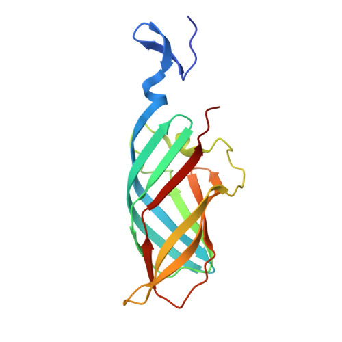
    Reovirus attachment protein sigma1 mediates engagement of receptors on the surface of target cells and undergoes dramatic conformational rearrangements during viral disassembly in the endocytic pathway. The sigma1 protein is a filamentous, trimeric molecule with a globular beta-barrel head domain. An unusual cluster of aspartic acid residues sandwiched between hydrophobic tyrosines is located at the sigma1 subunit interface. A 1.75-A structure of the sigma1 head domain now reveals two water molecules at the subunit interface that are held strictly in position and interact with neighboring residues. Structural and biochemical analyses of mutants affecting the aspartic acid sandwich indicate that these residues and the corresponding chelated water molecules act as a plug to block the free flow of solvent and stabilize the trimer. This arrangement of residues at the sigma1 head trimer interface illustrates a new protein design motif that may confer conformational mobility during cell entry.


  • Organizational Affiliation

    Interfakultäres Institut für Biochemie, Universität Tübingen, Hoppe-Seyler-Strasse 4, D-72076 Tübingen, Germany.


Macromolecules
Find similar proteins by:  (by identity cutoff)  |  3D Structure
Entity ID: 1
MoleculeChains Sequence LengthOrganismDetailsImage
Viral attachment protein sigma 1
A, B, C, D, E
A, B, C, D, E, F
165Reovirus sp.Mutation(s): 1 
Gene Names: S1
UniProt
Find proteins for P03528 (Reovirus type 3 (strain Dearing))
Explore P03528 
Go to UniProtKB:  P03528
Entity Groups  
Sequence Clusters30% Identity50% Identity70% Identity90% Identity95% Identity100% Identity
UniProt GroupP03528
Sequence Annotations
Expand
  • Reference Sequence
Experimental Data & Validation

Experimental Data

  • Method: X-RAY DIFFRACTION
  • Resolution: 1.85 Å
  • R-Value Free: 0.224 
  • R-Value Work: 0.175 
  • R-Value Observed: 0.180 
  • Space Group: P 1 21 1
Unit Cell:
Length ( Å )Angle ( ˚ )
a = 84.02α = 90
b = 51.6β = 95.59
c = 108.86γ = 90
Software Package:
Software NamePurpose
REFMACrefinement
MAR345data collection
DENZOdata reduction
SCALEPACKdata scaling
AMoREphasing

Structure Validation

View Full Validation Report


Entry History 

Deposition Data

Revision History  (Full details and data files)

  • Version 1.0: 2007-02-13
    Type: Initial release
  • Version 1.1: 2008-05-01
    Changes: Version format compliance
  • Version 1.2: 2011-07-13
    Changes: Version format compliance
  • Version 1.3: 2017-10-18
    Changes: Refinement description
  • Version 1.4: 2021-10-20
    Changes: Database references, Derived calculations
  • Version 1.5: 2023-08-30
    Changes: Data collection, Refinement description