2OEF

Open and Closed Structures of the UDP-Glucose Pyrophosphorylase from Leishmania major


Experimental Data Snapshot

  • Method: X-RAY DIFFRACTION
  • Resolution: 2.40 Å
  • R-Value Free: 0.261 
  • R-Value Work: 0.214 
  • R-Value Observed: 0.214 

wwPDB Validation   3D Report Full Report


This is version 1.3 of the entry. See complete history


Literature

Open and Closed Structures of the UDP-glucose Pyrophosphorylase from Leishmania major.

Steiner, T.Lamerz, A.C.Hess, P.Breithaupt, C.Krapp, S.Bourenkov, G.Huber, R.Gerardy-Schahn, R.Jacob, U.

(2007) J Biol Chem 282: 13003-13010

  • DOI: https://doi.org/10.1074/jbc.M609984200
  • Primary Citation of Related Structures:  
    2OEF, 2OEG

  • PubMed Abstract: 

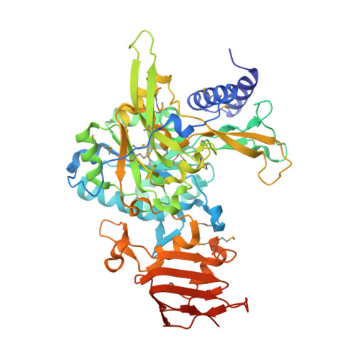
    Uridine diphosphate-glucose pyrophosphorylase (UGPase) represents a ubiquitous enzyme, which catalyzes the formation of UDP-glucose, a key metabolite of the carbohydrate pathways of all organisms. In the protozoan parasite Leishmania major, which causes a broad spectrum of diseases and is transmitted to humans by sand fly vectors, UGPase represents a virulence factor because of its requirement for the synthesis of cell surface glycoconjugates. Here we present the crystal structures of the L. major UGPase in its uncomplexed apo form (open conformation) and in complex with UDP-glucose (closed conformation). The UGPase consists of three distinct domains. The N-terminal domain exhibits species-specific differences in length, which might permit distinct regulation mechanisms. The central catalytic domain resembles a Rossmann-fold and contains key residues that are conserved in many nucleotidyltransferases. The C-terminal domain forms a left-handed parallel beta-helix (LbetaH), which represents a rarely observed structural element. The presented structures together with mutagenesis analyses provide a basis for a detailed analysis of the catalytic mechanism and for the design of species-specific UGPase inhibitors.


  • Organizational Affiliation

    Max-Planck-Institut für Biochemie, Abteilung für Strukturforschung, Am Klopferspitz 18, 82152 Martinsried, Germany. steiner@biochem.mpg.de


Macromolecules
Find similar proteins by:  (by identity cutoff)  |  3D Structure
Entity ID: 1
MoleculeChains Sequence LengthOrganismDetailsImage
UTP-glucose-1-phosphate uridylyltransferase 2, putative505Leishmania majorMutation(s): 18 
Gene Names: UGP
EC: 2.7.7.9
UniProt
Find proteins for Q4QDU3 (Leishmania major)
Explore Q4QDU3 
Go to UniProtKB:  Q4QDU3
Entity Groups  
Sequence Clusters30% Identity50% Identity70% Identity90% Identity95% Identity100% Identity
UniProt GroupQ4QDU3
Sequence Annotations
Expand
  • Reference Sequence
Small Molecules
Modified Residues  1 Unique
IDChains TypeFormula2D DiagramParent
MSE
Query on MSE
A
L-PEPTIDE LINKINGC5 H11 N O2 SeMET
Experimental Data & Validation

Experimental Data

  • Method: X-RAY DIFFRACTION
  • Resolution: 2.40 Å
  • R-Value Free: 0.261 
  • R-Value Work: 0.214 
  • R-Value Observed: 0.214 
  • Space Group: C 2 2 21
Unit Cell:
Length ( Å )Angle ( ˚ )
a = 72.952α = 90
b = 107.661β = 90
c = 150.131γ = 90
Software Package:
Software NamePurpose
CNSrefinement
DENZOdata reduction
SCALEPACKdata scaling
CNSphasing

Structure Validation

View Full Validation Report


Entry History 

Deposition Data

  • Released Date: 2007-02-13 
  • Deposition Author(s): Steiner, T.

Revision History  (Full details and data files)

  • Version 1.0: 2007-02-13
    Type: Initial release
  • Version 1.1: 2008-05-01
    Changes: Version format compliance
  • Version 1.2: 2011-07-13
    Changes: Version format compliance
  • Version 1.3: 2023-12-27
    Changes: Data collection, Database references, Derived calculations