2O6G

Crystal structure of IRF-3 bound to the interferon-b enhancer


Experimental Data Snapshot

  • Method: X-RAY DIFFRACTION
  • Resolution: 3.10 Å
  • R-Value Free: 0.265 
  • R-Value Work: 0.251 

wwPDB Validation   3D Report Full Report


This is version 1.4 of the entry. See complete history


Literature

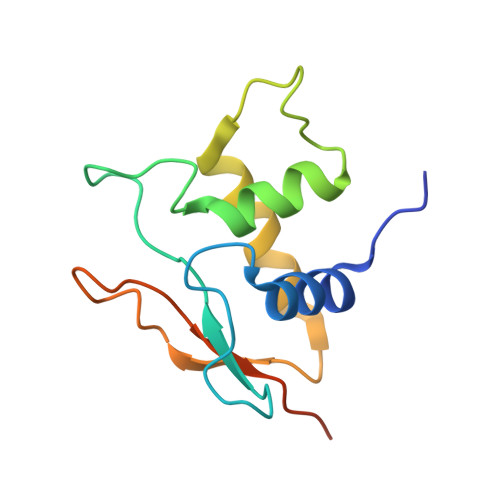
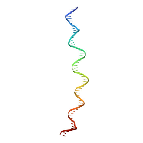
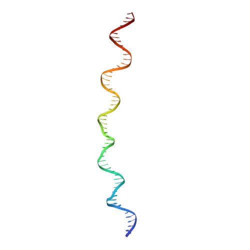
An Atomic Model of the Interferon-beta Enhanceosome.

Panne, D.Maniatis, T.Harrison, S.C.

(2007) Cell 129: 1111-1123

  • DOI: https://doi.org/10.1016/j.cell.2007.05.019
  • Primary Citation of Related Structures:  
    2O61, 2O6G

  • PubMed Abstract: 

    Transcriptional activation of the interferon-beta (IFN-beta) gene requires assembly of an enhanceosome containing ATF-2/c-Jun, IRF-3/IRF-7, and NFkappaB. These factors bind cooperatively to the IFN-beta enhancer and recruit coactivators and chromatin-remodeling proteins to the IFN-beta promoter. We describe here a crystal structure of the DNA-binding domains of IRF-3, IRF-7, and NFkappaB, bound to one half of the enhancer, and use a previously described structure of the remaining half to assemble a complete picture of enhanceosome architecture in the vicinity of the DNA. Association of eight proteins with the enhancer creates a continuous surface for recognizing a composite DNA-binding element. Paucity of local protein-protein contacts suggests that cooperative occupancy of the enhancer comes from both binding-induced changes in DNA conformation and interactions with additional components such as CBP. Contacts with virtually every nucleotide pair account for the evolutionary invariance of the enhancer sequence.


  • Organizational Affiliation

    The Jack and Eileen Connors Structural Biology Laboratory, Harvard Medical School, Department of Biological Chemistry and Molecular Pharmacology, Howard Hughes Medical Institute, 250 Longwood Avenue, Boston, MA 02115, USA.


Macromolecules

Find similar proteins by:  (by identity cutoff)  |  3D Structure
Entity ID: 3
MoleculeChains Sequence LengthOrganismDetailsImage
Interferon regulatory factor 3C [auth E],
D [auth F],
E [auth G],
F [auth H]
123Homo sapiensMutation(s): 0 
Gene Names: IRF3
UniProt & NIH Common Fund Data Resources
Find proteins for Q14653 (Homo sapiens)
Explore Q14653 
Go to UniProtKB:  Q14653
PHAROS:  Q14653
GTEx:  ENSG00000126456 
Entity Groups  
Sequence Clusters30% Identity50% Identity70% Identity90% Identity95% Identity100% Identity
UniProt GroupQ14653
Sequence Annotations
Expand
  • Reference Sequence
Find similar nucleic acids by:  (by identity cutoff)  |  3D Structure
Entity ID: 1
MoleculeChains LengthOrganismImage
interferon-b enhancerA [auth D]57N/A
Sequence Annotations
Expand
  • Reference Sequence
Find similar nucleic acids by:  (by identity cutoff)  |  3D Structure
Entity ID: 2
MoleculeChains LengthOrganismImage
interferon-b enhancerB [auth C]57N/A
Sequence Annotations
Expand
  • Reference Sequence
Experimental Data & Validation

Experimental Data

  • Method: X-RAY DIFFRACTION
  • Resolution: 3.10 Å
  • R-Value Free: 0.265 
  • R-Value Work: 0.251 
  • Space Group: I 2 2 2
Unit Cell:
Length ( Å )Angle ( ˚ )
a = 91.02α = 90
b = 105.32β = 90
c = 353.16γ = 90
Software Package:
Software NamePurpose
DENZOdata reduction
SCALEPACKdata scaling
CNSrefinement
PDB_EXTRACTdata extraction
ADSCdata collection
HKL-2000data reduction
MOLREPphasing

Structure Validation

View Full Validation Report


Entry History 

Deposition Data

  • Released Date: 2007-07-24 
  • Deposition Author(s): Panne, D.

Revision History  (Full details and data files)

  • Version 1.0: 2007-07-24
    Type: Initial release
  • Version 1.1: 2008-05-01
    Changes: Version format compliance
  • Version 1.2: 2011-07-13
    Changes: Version format compliance
  • Version 1.3: 2017-10-18
    Changes: Refinement description
  • Version 1.4: 2023-12-27
    Changes: Data collection, Database references