2O5X

Crystal structure of 1E9 LeuH47Trp/ArgH100Trp, an engineered Diels-Alderase Fab with nM steroid-binding affinity


Experimental Data Snapshot

  • Method: X-RAY DIFFRACTION
  • Resolution: 2.05 Å
  • R-Value Free: 0.204 
  • R-Value Work: 0.178 
  • R-Value Observed: 0.179 

wwPDB Validation   3D Report Full Report


This is version 1.2 of the entry. See complete history


Literature

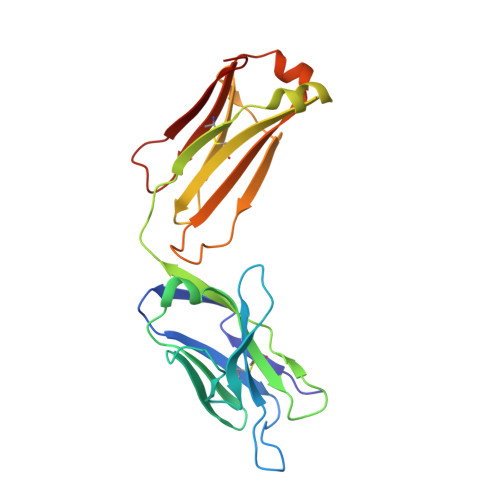
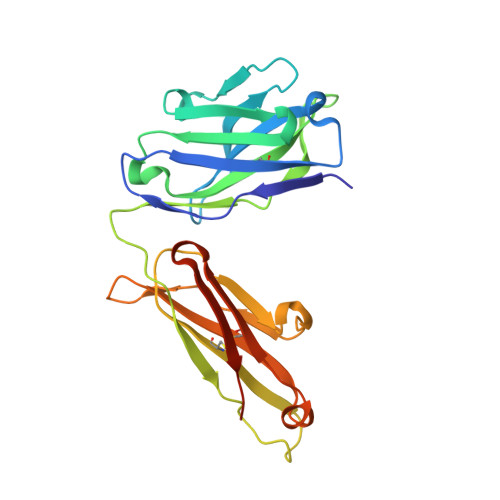
Closely related antibody receptors exploit fundamentally different strategies for steroid recognition.

Verdino, P.Aldag, C.Hilvert, D.Wilson, I.A.

(2008) Proc Natl Acad Sci U S A 105: 11725-11730

  • DOI: https://doi.org/10.1073/pnas.0801783105
  • Primary Citation of Related Structures:  
    2O5X, 2O5Y, 2O5Z

  • PubMed Abstract: 

    Molecular recognition by the adaptive immune system relies on specific high-affinity antibody receptors that are generated from a restricted set of starting sequences through homologous recombination and somatic mutation. The steroid binding antibody DB3 and the catalytic Diels-Alderase antibody 1E9 derive from the same germ line sequences but exhibit very distinct specificities and functions. However, mutation of only two of the 36 sequence differences in the variable domains, Leu(H47)Trp and Arg(H100)Trp, converts 1E9 into a high-affinity steroid receptor with a ligand recognition profile similar to DB3. To understand how these changes switch binding specificity and function, we determined the crystal structures of the 1E9 Leu(H47)Trp/Arg(H100)Trp double mutant (1E9dm) as an unliganded Fab at 2.05 A resolution and in complex with two configurationally distinct steroids at 2.40 and 2.85 A. Surprisingly, despite the functional mimicry of DB3, 1E9dm employs a distinct steroid binding mechanism. Extensive structural rearrangements occur in the combining site, where residue H47 acts as a specificity switch and H100 adapts to different ligands. Unlike DB3, 1E9dm does not use alternative binding pockets or different sets of hydrogen-bonding interactions to bind configurationally distinct steroids. Rather, the different steroids are inserted more deeply into the 1E9dm combining site, creating more hydrophobic contacts that energetically compensate for the lack of hydrogen bonds. These findings demonstrate how subtle mutations within an existing molecular scaffold can dramatically modulate the function of immune receptors by inducing unanticipated, but compensating, mechanisms of ligand interaction.


  • Organizational Affiliation

    Department of Molecular Biology and The Skaggs Institute for Chemical Biology, The Scripps Research Institute, 10550 North Torrey Pines Road, La Jolla, CA 92037, USA.


Macromolecules
Find similar proteins by:  (by identity cutoff)  |  3D Structure
Entity ID: 1
MoleculeChains Sequence LengthOrganismDetailsImage
chimeric antibody Fab 1E9-DB3A [auth L]219Mus musculusHomo sapiens
This entity is chimeric
Mutation(s): 0 
Entity Groups  
Sequence Clusters30% Identity50% Identity70% Identity90% Identity95% Identity100% Identity
Sequence Annotations
Expand
  • Reference Sequence
Find similar proteins by:  (by identity cutoff)  |  3D Structure
Entity ID: 2
MoleculeChains Sequence LengthOrganismDetailsImage
chimeric antibody Fab 1E9-DB3B [auth H]227Mus musculusHomo sapiens
This entity is chimeric
Mutation(s): 0 
Entity Groups  
Sequence Clusters30% Identity50% Identity70% Identity90% Identity95% Identity100% Identity
Sequence Annotations
Expand
  • Reference Sequence
Small Molecules
Ligands 2 Unique
IDChains Name / Formula / InChI Key2D Diagram3D Interactions
TRS
Query on TRS

Download Ideal Coordinates CCD File 
N [auth H]2-AMINO-2-HYDROXYMETHYL-PROPANE-1,3-DIOL
C4 H12 N O3
LENZDBCJOHFCAS-UHFFFAOYSA-O
SO4
Query on SO4

Download Ideal Coordinates CCD File 
C [auth L]
D [auth L]
E [auth L]
F [auth L]
G [auth L]
C [auth L],
D [auth L],
E [auth L],
F [auth L],
G [auth L],
H,
I [auth H],
J [auth H],
K [auth H],
L [auth H],
M [auth H]
SULFATE ION
O4 S
QAOWNCQODCNURD-UHFFFAOYSA-L
Experimental Data & Validation

Experimental Data

  • Method: X-RAY DIFFRACTION
  • Resolution: 2.05 Å
  • R-Value Free: 0.204 
  • R-Value Work: 0.178 
  • R-Value Observed: 0.179 
  • Space Group: P 31 2 1
Unit Cell:
Length ( Å )Angle ( ˚ )
a = 127.573α = 90
b = 127.573β = 90
c = 91.928γ = 120
Software Package:
Software NamePurpose
REFMACrefinement
BOSdata collection
HKL-2000data reduction
HKL-2000data scaling
PHASERphasing

Structure Validation

View Full Validation Report


Entry History 

Deposition Data

Revision History  (Full details and data files)

  • Version 1.0: 2007-12-18
    Type: Initial release
  • Version 1.1: 2011-07-13
    Changes: Advisory, Derived calculations, Version format compliance
  • Version 1.2: 2023-08-30
    Changes: Data collection, Database references, Derived calculations, Refinement description