Unique thermodynamic response of tipranavir to human immunodeficiency virus type 1 protease drug resistance mutations.
Muzammil, S., Armstrong, A.A., Kang, L.W., Jakalian, A., Bonneau, P.R., Schmelmer, V., Amzel, L.M., Freire, E.(2007) J Virol 81: 5144-5154
- PubMed: 17360759
- DOI: https://doi.org/10.1128/JVI.02706-06
- Primary Citation of Related Structures:
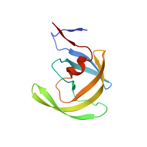
2O4K, 2O4L, 2O4N, 2O4P, 2O4S - PubMed Abstract:
Drug resistance is a major problem affecting the clinical efficacy of antiretroviral agents, including protease inhibitors, in the treatment of infection with human immunodeficiency virus type 1 (HIV-1)/AIDS. Consequently, the elucidation of the mechanisms by which HIV-1 protease inhibitors maintain antiviral activity in the presence of mutations is critical to the development of superior inhibitors. Tipranavir, a nonpeptidic HIV-1 protease inhibitor, has been recently approved for the treatment of HIV infection. Tipranavir inhibits wild-type protease with high potency (K(i) = 19 pM) and demonstrates durable efficacy in the treatment of patients infected with HIV-1 strains containing multiple common mutations associated with resistance. The high potency of tipranavir results from a very large favorable entropy change (-TDeltaS = -14.6 kcal/mol) combined with a favorable, albeit small, enthalpy change (DeltaH = -0.7 kcal/mol, 25 degrees C). Characterization of tipranavir binding to wild-type protease, active site mutants I50V and V82F/I84V, the multidrug-resistant mutant L10I/L33I/M46I/I54V/L63I/V82A/I84V/L90M, and the tipranavir in vitro-selected mutant I13V/V32L/L33F/K45I/V82L/I84V was performed by isothermal titration calorimetry and crystallography. Thermodynamically, the good response of tipranavir arises from a unique behavior: it compensates for entropic losses by actual enthalpic gains or by sustaining minimal enthalpic losses when facing the mutants. The net result is a small loss in binding affinity. Structurally, tipranavir establishes a very strong hydrogen bond network with invariant regions of the protease, which is maintained with the mutants, including catalytic Asp25 and the backbone of Asp29, Asp30, Gly48 and Ile50. Moreover, tipranavir forms hydrogen bonds directly to Ile50, while all other inhibitors do so by being mediated by a water molecule.
Organizational Affiliation:
Department of Biology, Johns Hopkins University, Baltimore, MD 21218, USA.