2NUO

Crystal structure of a complex of griffithsin with glucose


Experimental Data Snapshot

  • Method: X-RAY DIFFRACTION
  • Resolution: 1.50 Å
  • R-Value Free: 0.203 
  • R-Value Work: 0.155 
  • R-Value Observed: 0.157 

wwPDB Validation   3D Report Full Report


Ligand Structure Quality Assessment 


This is version 1.4 of the entry. See complete history


Literature

Crystallographic studies of the complexes of antiviral protein griffithsin with glucose and N-acetylglucosamine.

Ziolkowska, N.E.Shenoy, S.R.O'Keefe, B.R.Wlodawer, A.

(2007) Protein Sci 16: 1485-1489

  • DOI: https://doi.org/10.1110/ps.072889407
  • Primary Citation of Related Structures:  
    2NU5, 2NUO

  • PubMed Abstract: 

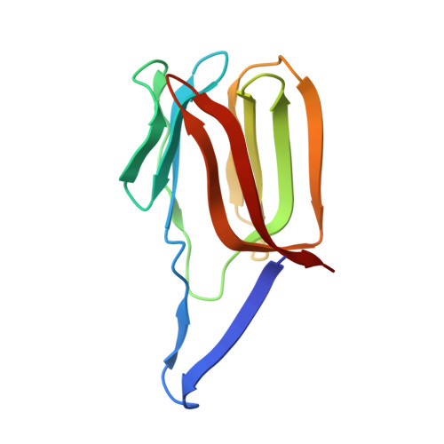
    Crystal structures of complexes of an antiviral lectin griffithsin (GRFT) with glucose and N-acetylglucosamine were solved and refined at high resolution. In both complexes, all six monosaccharide-binding sites of GRFT were occupied and the mode of binding was similar to that of mannose. In our previous attempts to obtain a complex with N-acetylglucosamine by soaking, only a single site was occupied; thus, cocrystallization was clearly superior despite lower concentration of the ligand. Isothermal titration calorimetric experiments with N-acetylglucosamine, glucose, and mannose provided enthalpic evidence of distinct binding differences between the three monosaccharides. A comparison of the mode of binding of different monosaccharides is discussed in the context of the antiviral activity of GRFT, based on specific binding to high-mannose-containing complex carbohydrates found on viral envelopes.


  • Organizational Affiliation

    Protein Structure Section, Macromolecular Crystallography Laboratory, National Cancer Institute, NCI-Frederick, MD 21702-1201, USA.


Macromolecules
Find similar proteins by:  (by identity cutoff)  |  3D Structure
Entity ID: 1
MoleculeChains Sequence LengthOrganismDetailsImage
Griffithsin
A, B
122GriffithsiaMutation(s): 1 
UniProt
Find proteins for P84801 (Griffithsia sp. (strain Q66D336))
Explore P84801 
Go to UniProtKB:  P84801
Entity Groups  
Sequence Clusters30% Identity50% Identity70% Identity90% Identity95% Identity100% Identity
UniProt GroupP84801
Sequence Annotations
Expand
  • Reference Sequence
Small Molecules
Ligands 3 Unique
IDChains Name / Formula / InChI Key2D Diagram3D Interactions
BGC
Query on BGC

Download Ideal Coordinates CCD File 
C [auth A]
D [auth A]
E [auth A]
H [auth B]
I [auth B]
C [auth A],
D [auth A],
E [auth A],
H [auth B],
I [auth B],
J [auth B]
beta-D-glucopyranose
C6 H12 O6
WQZGKKKJIJFFOK-VFUOTHLCSA-N
SO4
Query on SO4

Download Ideal Coordinates CCD File 
F [auth A]SULFATE ION
O4 S
QAOWNCQODCNURD-UHFFFAOYSA-L
EDO
Query on EDO

Download Ideal Coordinates CCD File 
G [auth A]1,2-ETHANEDIOL
C2 H6 O2
LYCAIKOWRPUZTN-UHFFFAOYSA-N
Experimental Data & Validation

Experimental Data

  • Method: X-RAY DIFFRACTION
  • Resolution: 1.50 Å
  • R-Value Free: 0.203 
  • R-Value Work: 0.155 
  • R-Value Observed: 0.157 
  • Space Group: P 21 21 21
Unit Cell:
Length ( Å )Angle ( ˚ )
a = 37.49α = 90
b = 53.67β = 90
c = 103.83γ = 90
Software Package:
Software NamePurpose
REFMACrefinement
MAR345data collection
DENZOdata reduction
SCALEPACKdata scaling
PHASERphasing

Structure Validation

View Full Validation Report


Ligand Structure Quality Assessment 


Entry History 

Deposition Data

Revision History  (Full details and data files)

  • Version 1.0: 2007-08-07
    Type: Initial release
  • Version 1.1: 2011-07-13
    Changes: Advisory, Derived calculations, Version format compliance
  • Version 1.2: 2017-10-18
    Changes: Advisory, Refinement description
  • Version 1.3: 2020-07-29
    Type: Remediation
    Reason: Carbohydrate remediation
    Changes: Advisory, Data collection, Database references, Derived calculations, Structure summary
  • Version 1.4: 2023-08-30
    Changes: Advisory, Data collection, Database references, Refinement description, Structure summary