2NQJ

Crystal structure of Escherichia coli endonuclease IV (Endo IV) E261Q mutant bound to damaged DNA


Experimental Data Snapshot

  • Method: X-RAY DIFFRACTION
  • Resolution: 2.45 Å
  • R-Value Free: 0.254 
  • R-Value Work: 0.198 

wwPDB Validation   3D Report Full Report


This is version 1.3 of the entry. See complete history


Literature

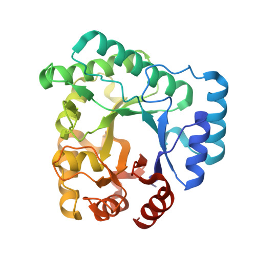
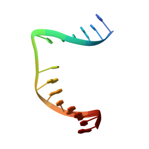
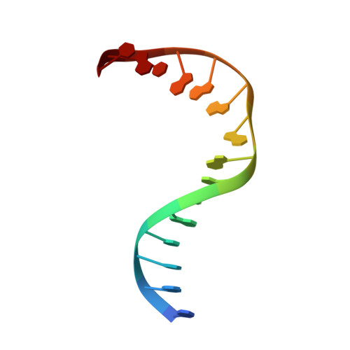
DNA apurinic-apyrimidinic site binding and excision by endonuclease IV.

Garcin, E.D.Hosfield, D.J.Desai, S.A.Haas, B.J.Bjoras, M.Cunningham, R.P.Tainer, J.A.

(2008) Nat Struct Mol Biol 15: 515-522

  • DOI: https://doi.org/10.1038/nsmb.1414
  • Primary Citation of Related Structures:  
    2NQ9, 2NQH, 2NQJ

  • PubMed Abstract: 

    Escherichia coli endonuclease IV is an archetype for an abasic or apurinic-apyrimidinic endonuclease superfamily crucial for DNA base excision repair. Here biochemical, mutational and crystallographic characterizations reveal a three-metal ion mechanism for damage binding and incision. The 1.10-A resolution DNA-free and the 2.45-A resolution DNA-substrate complex structures capture substrate stabilization by Arg37 and reveal a distorted Zn3-ligand arrangement that reverts, after catalysis, to an ideal geometry suitable to hold rather than release cleaved DNA product. The 1.45-A resolution DNA-product complex structure shows how Tyr72 caps the active site, tunes its dielectric environment and promotes catalysis by Glu261-activated hydroxide, bound to two Zn2+ ions throughout catalysis. These structural, mutagenesis and biochemical results suggest general requirements for abasic site removal in contrast to features specific to the distinct endonuclease IV alpha-beta triose phosphate isomerase (TIM) barrel and APE1 four-layer alpha-beta folds of the apurinic-apyrimidinic endonuclease families.


  • Organizational Affiliation

    Department of Molecular Biology and Skaggs Institute for Chemical Biology, The Scripps Research Institute, 10550 North Torrey Pines Road, MB4 La Jolla, California 92037, USA.


Macromolecules

Find similar proteins by:  (by identity cutoff)  |  3D Structure
Entity ID: 3
MoleculeChains Sequence LengthOrganismDetailsImage
Endonuclease 4C [auth A],
D [auth B]
285Escherichia coliMutation(s): 1 
Gene Names: nfo
EC: 3.1.21.2
UniProt
Find proteins for P0A6C1 (Escherichia coli (strain K12))
Explore P0A6C1 
Go to UniProtKB:  P0A6C1
Entity Groups  
Sequence Clusters30% Identity50% Identity70% Identity90% Identity95% Identity100% Identity
UniProt GroupP0A6C1
Sequence Annotations
Expand
  • Reference Sequence

Find similar nucleic acids by:  Sequence   |   3D Structure  

Entity ID: 1
MoleculeChains LengthOrganismImage
5'-D(*GP*CP*GP*TP*CP*CP*(3DR)P*CP*GP*AP*CP*GP*AP*CP*G)-3'A [auth C]15N/A
Sequence Annotations
Expand
  • Reference Sequence

Find similar nucleic acids by:  Sequence   |   3D Structure  

Entity ID: 2
MoleculeChains LengthOrganismImage
5'-D(*CP*GP*TP*CP*GP*TP*CP*GP*GP*GP*GP*AP*CP*GP*C)-3'B [auth D]15N/A
Sequence Annotations
Expand
  • Reference Sequence
Experimental Data & Validation

Experimental Data

  • Method: X-RAY DIFFRACTION
  • Resolution: 2.45 Å
  • R-Value Free: 0.254 
  • R-Value Work: 0.198 
  • Space Group: P 21 21 21
Unit Cell:
Length ( Å )Angle ( ˚ )
a = 55.6α = 90
b = 86.2β = 90
c = 148.3γ = 90
Software Package:
Software NamePurpose
CNSrefinement
PDB_EXTRACTdata extraction
DENZOdata reduction
SCALEPACKdata scaling
EPMRphasing

Structure Validation

View Full Validation Report


Entry History 

Deposition Data

Revision History  (Full details and data files)

  • Version 1.0: 2007-11-06
    Type: Initial release
  • Version 1.1: 2011-07-13
    Changes: Version format compliance
  • Version 1.2: 2021-10-20
    Changes: Database references, Derived calculations
  • Version 1.3: 2023-08-30
    Changes: Data collection, Refinement description