2NLL

RETINOID X RECEPTOR-THYROID HORMONE RECEPTOR DNA-BINDING DOMAIN HETERODIMER BOUND TO THYROID RESPONSE ELEMENT DNA


Experimental Data Snapshot

  • Method: X-RAY DIFFRACTION
  • Resolution: 1.90 Å
  • R-Value Free: 0.269 
  • R-Value Work: 0.206 
  • R-Value Observed: 0.206 

wwPDB Validation   3D Report Full Report


This is version 1.3 of the entry. See complete history


Literature

Structural determinants of nuclear receptor assembly on DNA direct repeats.

Rastinejad, F.Perlmann, T.Evans, R.M.Sigler, P.B.

(1995) Nature 375: 203-211

  • DOI: https://doi.org/10.1038/375203a0
  • Primary Citation of Related Structures:  
    2NLL

  • PubMed Abstract: 

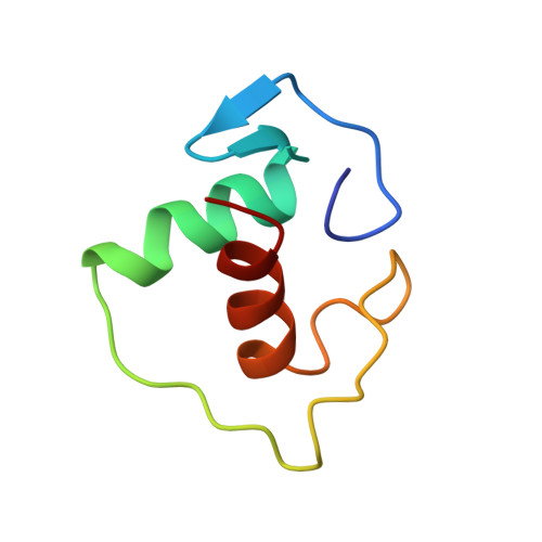
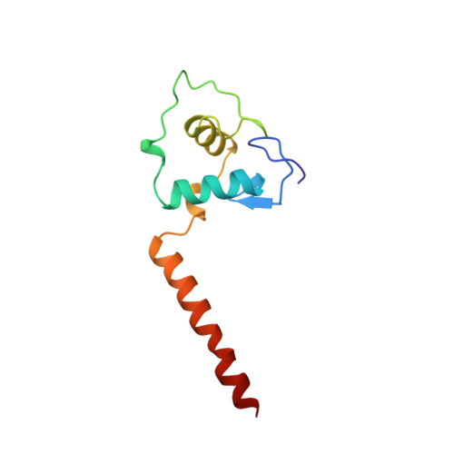
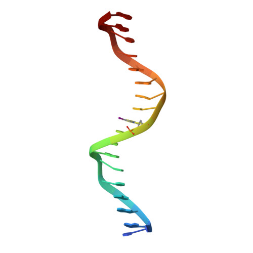
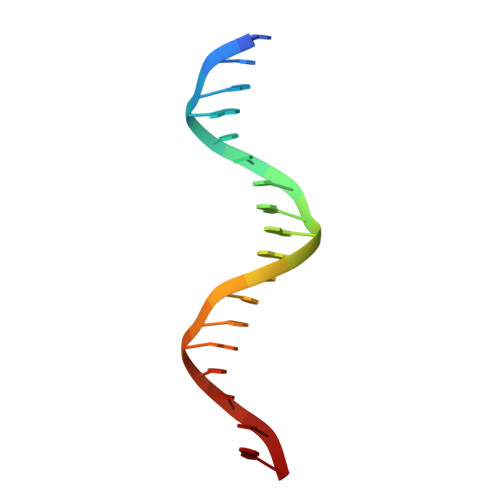
    Nuclear receptor heterodimers recognize response elements composed of two direct repeats of the consensus sequence 5'-AGGTCA-3' separated by one to five base pairs. The 1.9 A crystal structure of the complex formed by the DNA-binding domains of the 9-cis retinoic acid receptor and thyroid hormone receptor bound to a thyroid-response element shows that the subunits interact through a DNA-supported interface involving the carboxy-terminal extension of the DNA-binding domain of the thyroid hormone receptor. The stereochemistry suggests a mechanism by which heterodimers recognize the inter-half-site spacing between direct repeats.


  • Organizational Affiliation

    Department of Molecular Biophysics and Biochemistry, Yale University, New Haven, Connecticut 06510, USA.


Macromolecules

Find similar proteins by:  (by identity cutoff)  |  3D Structure
Entity ID: 3
MoleculeChains Sequence LengthOrganismDetailsImage
PROTEIN (RETINOIC ACID RECEPTOR)C [auth A]66Homo sapiensMutation(s): 0 
UniProt & NIH Common Fund Data Resources
Find proteins for P19793 (Homo sapiens)
Explore P19793 
Go to UniProtKB:  P19793
PHAROS:  P19793
GTEx:  ENSG00000186350 
Entity Groups  
Sequence Clusters30% Identity50% Identity70% Identity90% Identity95% Identity100% Identity
UniProt GroupP19793
Sequence Annotations
Expand
  • Reference Sequence
Find similar proteins by:  (by identity cutoff)  |  3D Structure
Entity ID: 4
MoleculeChains Sequence LengthOrganismDetailsImage
PROTEIN (THYROID HORMONE RECEPTOR)D [auth B]103Homo sapiensMutation(s): 0 
UniProt & NIH Common Fund Data Resources
Find proteins for P10828 (Homo sapiens)
Explore P10828 
Go to UniProtKB:  P10828
PHAROS:  P10828
GTEx:  ENSG00000151090 
Entity Groups  
Sequence Clusters30% Identity50% Identity70% Identity90% Identity95% Identity100% Identity
UniProt GroupP10828
Sequence Annotations
Expand
  • Reference Sequence

Find similar nucleic acids by:  Sequence   |   3D Structure  

Entity ID: 1
MoleculeChains LengthOrganismImage
DNA (5'-D(*CP*AP*GP*GP*TP*CP*AP*TP*TP*(5IU)P*CP*AP*GP*GP*TP*CP*AP*G)-3')A [auth C]18N/A
Sequence Annotations
Expand
  • Reference Sequence

Find similar nucleic acids by:  Sequence   |   3D Structure  

Entity ID: 2
MoleculeChains LengthOrganismImage
DNA (5'-D(*CP*TP*GP*AP*CP*CP*TP*GP*AP*AP*AP*TP*GP*AP*CP*CP*T P*G)-3')B [auth D]18N/A
Sequence Annotations
Expand
  • Reference Sequence
Experimental Data & Validation

Experimental Data

  • Method: X-RAY DIFFRACTION
  • Resolution: 1.90 Å
  • R-Value Free: 0.269 
  • R-Value Work: 0.206 
  • R-Value Observed: 0.206 
  • Space Group: P 21 21 21
Unit Cell:
Length ( Å )Angle ( ˚ )
a = 40.9α = 90
b = 65.6β = 90
c = 125.7γ = 90
Software Package:
Software NamePurpose
MLPHAREphasing
X-PLORrefinement
DENZOdata reduction
SCALEPACKdata scaling

Structure Validation

View Full Validation Report


Entry History 

Deposition Data

Revision History  (Full details and data files)

  • Version 1.0: 1997-03-12
    Type: Initial release
  • Version 1.1: 2008-05-22
    Changes: Version format compliance
  • Version 1.2: 2011-07-13
    Changes: Version format compliance
  • Version 1.3: 2024-02-21
    Changes: Data collection, Database references, Derived calculations