2MIP

CRYSTAL STRUCTURE OF HUMAN IMMUNODEFICIENCY VIRUS (HIV) TYPE 2 PROTEASE IN COMPLEX WITH A REDUCED AMIDE INHIBITOR AND COMPARISON WITH HIV-1 PROTEASE STRUCTURES


Experimental Data Snapshot

  • Method: X-RAY DIFFRACTION
  • Resolution: 2.20 Å
  • R-Value Work: 0.176 
  • R-Value Observed: 0.176 

wwPDB Validation   3D Report Full Report


This is version 1.4 of the entry. See complete history


Literature

Crystal structure of human immunodeficiency virus (HIV) type 2 protease in complex with a reduced amide inhibitor and comparison with HIV-1 protease structures.

Tong, L.Pav, S.Pargellis, C.Do, F.Lamarre, D.Anderson, P.C.

(1993) Proc Natl Acad Sci U S A 90: 8387-8391

  • DOI: https://doi.org/10.1073/pnas.90.18.8387
  • Primary Citation of Related Structures:  
    2MIP

  • PubMed Abstract: 

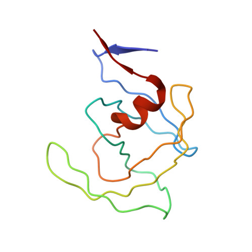
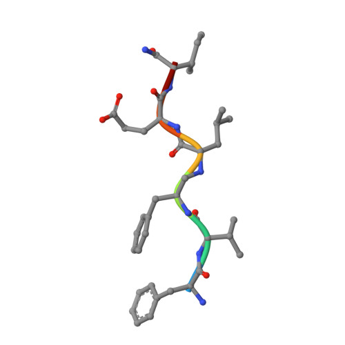
    The crystal structure of HIV-2 protease in complex with a reduced amide inhibitor [BI-LA-398; Phe-Val-Phe-psi (CH2NH)-Leu-Glu-Ile-amide] has been determined at 2.2-A resolution and refined to a crystallographic R factor of 17.6%. The rms deviation from ideality in bond lengths is 0.018 A and in bond angles is 2.8 degrees. The largest structural differences between HIV-1 and HIV-2 proteases are located at residues 15-20, 34-40, and 65-73, away from the flap region and the substrate binding sites. The rms distance between equivalent C alpha atoms of HIV-1 and HIV-2 protease structures excluding these residues is 0.5 A. The shapes of the S1 and S2 pockets in the presence of this inhibitor are essentially unperturbed by the amino acid differences between HIV-1 and HIV-2 proteases. The interaction of the inhibitor with HIV-2 protease is similar to that observed in HIV-1 protease structures. The unprotected N terminus of the inhibitor interacts with the side chains of Asp-29 and Asp-30. The glutamate side chain of the inhibitor forms hydrogen bonds with the main-chain amido groups of residues 129 and 130.


  • Organizational Affiliation

    Department of Medicinal Chemistry, Boehringer Ingelheim Pharmaceutical, Inc., Ridgefield, CT 06877.


Macromolecules
Find similar proteins by:  (by identity cutoff)  |  3D Structure
Entity ID: 1
MoleculeChains Sequence LengthOrganismDetailsImage
HIV-2 PROTEASE
A, B, C, D
99Human immunodeficiency virus 2Mutation(s): 0 
UniProt
Find proteins for P04584 (Human immunodeficiency virus type 2 subtype A (isolate ROD))
Explore P04584 
Go to UniProtKB:  P04584
Entity Groups  
Sequence Clusters30% Identity50% Identity70% Identity90% Identity95% Identity100% Identity
UniProt GroupP04584
Sequence Annotations
Expand
  • Reference Sequence

Find similar proteins by:  Sequence   |   3D Structure  

Entity ID: 2
MoleculeChains Sequence LengthOrganismDetailsImage
INHIBITOR BI-LA-398
E, F, G, H
7N/AMutation(s): 0 
Sequence Annotations
Expand
  • Reference Sequence
Experimental Data & Validation

Experimental Data

  • Method: X-RAY DIFFRACTION
  • Resolution: 2.20 Å
  • R-Value Work: 0.176 
  • R-Value Observed: 0.176 
  • Space Group: P 43
Unit Cell:
Length ( Å )Angle ( ˚ )
a = 55.1α = 90
b = 55.1β = 90
c = 138.9γ = 90
Software Package:
Software NamePurpose
X-PLORmodel building
TNTrefinement
X-PLORrefinement
X-PLORphasing

Structure Validation

View Full Validation Report


Entry History 

Deposition Data

Revision History  (Full details and data files)

  • Version 1.0: 1993-10-31
    Type: Initial release
  • Version 1.1: 2008-03-24
    Changes: Version format compliance
  • Version 1.2: 2011-07-13
    Changes: Atomic model, Database references, Derived calculations, Non-polymer description, Structure summary, Version format compliance
  • Version 1.3: 2019-07-17
    Changes: Data collection, Derived calculations, Other, Refinement description
  • Version 1.4: 2019-08-14
    Changes: Data collection, Refinement description