2KGX

HADDOCK structure of the talin F3 domain in complex with talin 1655-1822


Experimental Data Snapshot

  • Method: SOLUTION NMR
  • Conformers Calculated: 200 
  • Conformers Submitted: 
  • Selection Criteria: structures with the lowest energy 

wwPDB Validation   3D Report Full Report

Currently 2KGX does not have a validation slider image.


This is version 1.2 of the entry. See complete history


Literature

The structure of an interdomain complex that regulates talin activity.

Goult, B.T.Bate, N.Anthis, N.J.Wegener, K.L.Gingras, A.R.Patel, B.Barsukov, I.L.Campbell, I.D.Roberts, G.C.Critchley, D.R.

(2009) J Biol Chem 284: 15097-15106

  • DOI: https://doi.org/10.1074/jbc.M900078200
  • Primary Citation of Related Structures:  
    2KBB, 2KGX

  • PubMed Abstract: 

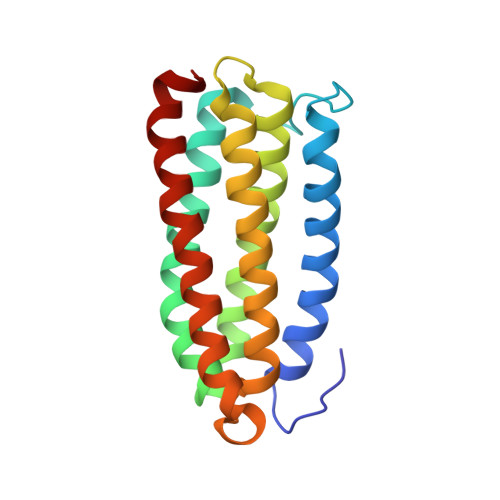
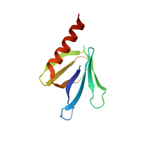
    Talin is a large flexible rod-shaped protein that activates the integrin family of cell adhesion molecules and couples them to cytoskeletal actin. It exists in both globular and extended conformations, and an intramolecular interaction between the N-terminal F3 FERM subdomain and the C-terminal part of the talin rod contributes to an autoinhibited form of the molecule. Here, we report the solution structure of the primary F3 binding domain within the C-terminal region of the talin rod and use intermolecular nuclear Overhauser effects to determine the structure of the complex. The rod domain (residues 1655-1822) is an amphipathic five-helix bundle; Tyr-377 of F3 docks into a hydrophobic pocket at one end of the bundle, whereas a basic loop in F3 (residues 316-326) interacts with a cluster of acidic residues in the middle of helix 4. Mutation of Glu-1770 abolishes binding. The rod domain competes with beta3-integrin tails for binding to F3, and the structure of the complex suggests that the rod is also likely to sterically inhibit binding of the FERM domain to the membrane.


  • Organizational Affiliation

    Department of Biochemistry, University of Leicester, Lancaster Road, Leicester LE1 9HN, UK.


Macromolecules
Find similar proteins by:  (by identity cutoff)  |  3D Structure
Entity ID: 1
MoleculeChains Sequence LengthOrganismDetailsImage
Talin-1174Mus musculusMutation(s): 0 
Gene Names: Tln1Tln
UniProt
Find proteins for P26039 (Mus musculus)
Explore P26039 
Go to UniProtKB:  P26039
Entity Groups  
Sequence Clusters30% Identity50% Identity70% Identity90% Identity95% Identity100% Identity
UniProt GroupP26039
Sequence Annotations
Expand
  • Reference Sequence
Find similar proteins by:  (by identity cutoff)  |  3D Structure
Entity ID: 2
MoleculeChains Sequence LengthOrganismDetailsImage
MKIAA1027 protein91Mus musculusMutation(s): 1 
Gene Names: Tln1mKIAA1027
UniProt
Find proteins for P26039 (Mus musculus)
Explore P26039 
Go to UniProtKB:  P26039
Entity Groups  
Sequence Clusters30% Identity50% Identity70% Identity90% Identity95% Identity100% Identity
UniProt GroupP26039
Sequence Annotations
Expand
  • Reference Sequence
Experimental Data & Validation

Experimental Data

  • Method: SOLUTION NMR
  • Conformers Calculated: 200 
  • Conformers Submitted: 
  • Selection Criteria: structures with the lowest energy 

Structure Validation

View Full Validation Report

Currently 2KGX does not have a validation slider image.


Entry History 

Deposition Data

Revision History  (Full details and data files)

  • Version 1.0: 2009-03-31
    Type: Initial release
  • Version 1.1: 2011-07-13
    Changes: Version format compliance
  • Version 1.2: 2021-10-20
    Changes: Database references, Derived calculations