2JJI

Endothiapepsin in complex with a gem-diol inhibitor.


Experimental Data Snapshot

  • Method: X-RAY DIFFRACTION
  • Resolution: 1.57 Å
  • R-Value Free: 0.196 
  • R-Value Observed: 0.157 

wwPDB Validation   3D Report Full Report


Ligand Structure Quality Assessment 


This is version 2.0 of the entry. See complete history


Literature

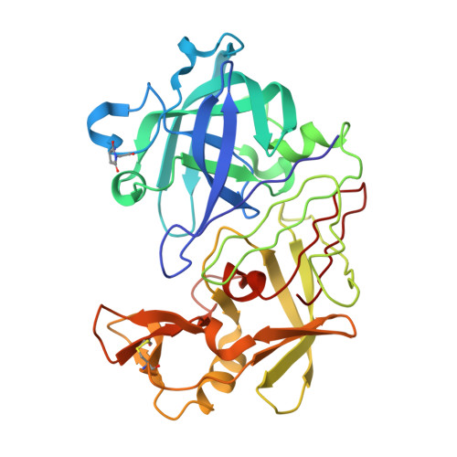
The Catalytic Mechanism of an Aspartic Proteinase Explored with Neutron and X-Ray Diffraction

Coates, L.Tuan, H.-F.Tomanicek, S.J.Kovalevsky, A.Mustyakimov, M.Erskine, P.Cooper, J.

(2008) J Am Chem Soc 130: 7235

  • DOI: https://doi.org/10.1021/ja801269x
  • Primary Citation of Related Structures:  
    2JJI, 2JJJ, 2VS2

  • PubMed Abstract: 

    Hydrogen atoms play key roles in enzyme mechanism, but as this study shows, even high-quality X-ray data to a resolution of 1 A cannot directly visualize them. Neutron diffraction, however, can locate deuterium atoms even at resolutions around 2 A. Both neutron and X-ray diffraction data have been used to investigate the transition state of the aspartic proteinase endothiapepsin. The different techniques reveal a different part of the story, revealing the clearest picture yet of the catalytic mechanism by which the enzyme operates. Room temperature neutron and X-ray diffraction data were used in a newly developed joint refinement software package to visualize deuterium atoms within the active site of the enzyme when a gem-diol transition state analogue inhibitor is bound at the active site. These data were also used to estimate their individual occupancy, while analysis of the differences between the bond lengths of the catalytic aspartates was performed using atomic resolution X-ray data. The two methods are in agreement on the protonation state of the active site with a transition state analogue inhibitor bound confirming the catalytic mechanism at which the enzyme operates.


  • Organizational Affiliation

    Spallation Neutron Source, Oak Ridge National Laboratory, 1 Bethel Valley Road, Oak Ridge, Tennessee 37831, USA.


Macromolecules
Find similar proteins by:  (by identity cutoff)  |  3D Structure
Entity ID: 1
MoleculeChains Sequence LengthOrganismDetailsImage
ENDOTHIAPEPSIN329Cryphonectria parasiticaMutation(s): 0 
EC: 3.4.23.22
UniProt
Find proteins for P11838 (Cryphonectria parasitica)
Explore P11838 
Go to UniProtKB:  P11838
Entity Groups  
Sequence Clusters30% Identity50% Identity70% Identity90% Identity95% Identity100% Identity
UniProt GroupP11838
Sequence Annotations
Expand
  • Reference Sequence
Small Molecules
Ligands 2 Unique
IDChains Name / Formula / InChI Key2D Diagram3D Interactions
0QS
Query on 0QS

Download Ideal Coordinates CCD File 
B [auth A]N~2~-[(2R)-2-benzyl-3-(tert-butylsulfonyl)propanoyl]-N-{(1R)-1-(cyclohexylmethyl)-3,3-difluoro-2,2-dihydroxy-4-[(2-morpholin-4-ylethyl)amino]-4-oxobutyl}-3-(1H-imidazol-3-ium-4-yl)-L-alaninamide
C36 H56 F2 N7 O8 S
RRJUDVVMLCYZDV-DTXPUJKBSA-O
SO4
Query on SO4

Download Ideal Coordinates CCD File 
C [auth A]SULFATE ION
O4 S
QAOWNCQODCNURD-UHFFFAOYSA-L
Modified Residues  1 Unique
IDChains TypeFormula2D DiagramParent
SUI
Query on SUI
A
L-PEPTIDE LINKINGC6 H8 N2 O4ASP, GLY
Biologically Interesting Molecules (External Reference) 1 Unique
Experimental Data & Validation

Experimental Data

  • Method: X-RAY DIFFRACTION
  • Resolution: 1.57 Å
  • R-Value Free: 0.196 
  • R-Value Observed: 0.157 
  • Space Group: P 1 21 1
Unit Cell:
Length ( Å )Angle ( ˚ )
a = 43.03α = 90
b = 75.72β = 97.02
c = 42.97γ = 90
Software Package:
Software NamePurpose
SHELXL-97refinement

Structure Validation

View Full Validation Report


Ligand Structure Quality Assessment 


Entry History 

Deposition Data

Revision History  (Full details and data files)

  • Version 1.0: 2008-05-27
    Type: Initial release
  • Version 1.1: 2011-07-13
    Changes: Atomic model, Database references, Derived calculations, Non-polymer description, Structure summary, Version format compliance
  • Version 1.2: 2013-03-06
    Changes: Other
  • Version 1.3: 2013-09-25
    Changes: Database references, Other
  • Version 1.4: 2018-04-04
    Changes: Data collection
  • Version 1.5: 2019-05-22
    Changes: Data collection, Derived calculations, Refinement description
  • Version 2.0: 2023-11-15
    Changes: Atomic model, Data collection, Database references, Derived calculations, Other