2JGP

Structure of the TycC5-6 PCP-C bidomain of the tyrocidine synthetase TycC


Experimental Data Snapshot

  • Method: X-RAY DIFFRACTION
  • Resolution: 1.85 Å
  • R-Value Free: 0.238 
  • R-Value Work: 0.208 
  • R-Value Observed: 0.209 

wwPDB Validation   3D Report Full Report


This is version 1.3 of the entry. See complete history


Literature

Structural and Functional Insights Into a Peptide Bond-Forming Bidomain from a Nonribosomal Peptide Synthetase.

Samel, S.A.Schoenafinger, G.Knappe, T.A.Marahiel, M.A.Essen, L.-O.

(2007) Structure 15: 781

  • DOI: https://doi.org/10.1016/j.str.2007.05.008
  • Primary Citation of Related Structures:  
    2JGP

  • PubMed Abstract: 

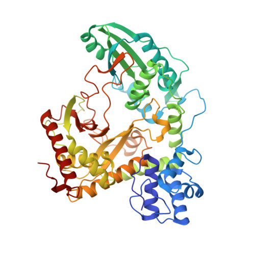
    The crystal structure of the bidomain PCP-C from modules 5 and 6 of the nonribosomal tyrocidine synthetase TycC was determined at 1.8 A resolution. The bidomain structure reveals a V-shaped condensation domain, the canyon-like active site groove of which is associated with the preceding peptidyl carrier protein (PCP) domain at its donor side. The relative arrangement of the PCP and the peptide bond-forming condensation (C) domain places the active sites approximately 50 A apart. Accordingly, this PCP-C structure represents a conformational state prior to peptide transfer from the donor-PCP to the acceptor-PCP domain, implying the existence of additional states of PCP-C domain interaction during catalysis. Additionally, PCP-C exerts a mode of cyclization activity that mimics peptide bond formation catalyzed by C domains. Based on mutational data and pK value analysis of active site residues, it is suggested that nonribosomal peptide bond formation depends on electrostatic interactions rather than on general acid/base catalysis.


  • Organizational Affiliation

    Department of Chemistry/Biochemistry, Philipps-Universität, Hans-Meerwein-Strasse, D-35032 Marburg, Germany.


Macromolecules
Find similar proteins by:  (by identity cutoff)  |  3D Structure
Entity ID: 1
MoleculeChains Sequence LengthOrganismDetailsImage
TYROCIDINE SYNTHETASE 3520Brevibacillus brevisMutation(s): 0 
UniProt
Find proteins for O30409 (Brevibacillus parabrevis)
Explore O30409 
Go to UniProtKB:  O30409
Entity Groups  
Sequence Clusters30% Identity50% Identity70% Identity90% Identity95% Identity100% Identity
UniProt GroupO30409
Sequence Annotations
Expand
  • Reference Sequence
Experimental Data & Validation

Experimental Data

  • Method: X-RAY DIFFRACTION
  • Resolution: 1.85 Å
  • R-Value Free: 0.238 
  • R-Value Work: 0.208 
  • R-Value Observed: 0.209 
  • Space Group: P 43 21 2
Unit Cell:
Length ( Å )Angle ( ˚ )
a = 84.973α = 90
b = 84.973β = 90
c = 164.969γ = 90
Software Package:
Software NamePurpose
MOSFLMdata reduction
SCALAdata scaling
SHELXphasing
SHARPphasing
REFMACrefinement

Structure Validation

View Full Validation Report


Entry History 

Deposition Data

Revision History  (Full details and data files)

  • Version 1.0: 2007-10-30
    Type: Initial release
  • Version 1.1: 2011-07-13
    Changes: Advisory, Version format compliance
  • Version 1.2: 2012-06-13
    Changes: Other
  • Version 1.3: 2019-07-24
    Changes: Advisory, Data collection