2JET

Crystal structure of a trypsin-like mutant (S189D , A226G) chymotrypsin.


Experimental Data Snapshot

  • Method: X-RAY DIFFRACTION
  • Resolution: 2.20 Å
  • R-Value Free: 0.334 
  • R-Value Work: 0.276 
  • R-Value Observed: 0.279 

wwPDB Validation   3D Report Full Report


This is version 1.4 of the entry. See complete history


Literature

The Crystal Structure of a Trypsin-Like Mutant Chymotrypsin: The Role of Position 226 in the Activity and Specificity of S189D Chymotrypsin.

Jelinek, B.Katona, G.Fodor, K.Venekei, I.Graf, L.

(2008) Protein J 27: 79

  • DOI: https://doi.org/10.1007/s10930-007-9110-3
  • Primary Citation of Related Structures:  
    2JET

  • PubMed Abstract: 

    The crystal structure of the S189D+A226G rat chymotrypsin-B mutant has been determined at 2.2 angstroms resolution. This mutant is the most trypsin-like mutant so far in the line of chymotrypsin-to-trypsin conversions, aiming for a more complete understanding of the structural basis of substrate specificity in pancreatic serine proteases. A226G caused significant rearrangements relative to S189D chymotrypsin, allowing an internal conformation of Asp189 which is close to that in trypsin. Serious distortions remain, however, in the activation domain, including zymogen-like features. The pH-profile of activity suggests that the conformation of the S1-site of the mutant is influenced also by the P1 residue of the substrate.


  • Organizational Affiliation

    Department of Biochemistry, Eötvös Loránd University, Pázmány s. 1/C, Budapest, 1117, Hungary. jelinek@elte.hu


Macromolecules

Find similar proteins by:  Sequence   |   3D Structure  

Entity ID: 1
MoleculeChains Sequence LengthOrganismDetailsImage
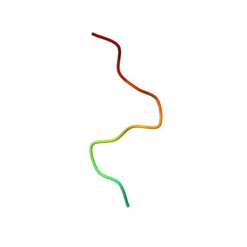
CHYMOTRYPSINOGEN B CHAIN A15Rattus norvegicusMutation(s): 0 
EC: 3.4.21.1
UniProt
Find proteins for P07338 (Rattus norvegicus)
Explore P07338 
Go to UniProtKB:  P07338
Entity Groups  
Sequence Clusters30% Identity50% Identity70% Identity90% Identity95% Identity100% Identity
UniProt GroupP07338
Sequence Annotations
Expand
  • Reference Sequence
Find similar proteins by:  (by identity cutoff)  |  3D Structure
Entity ID: 2
MoleculeChains Sequence LengthOrganismDetailsImage
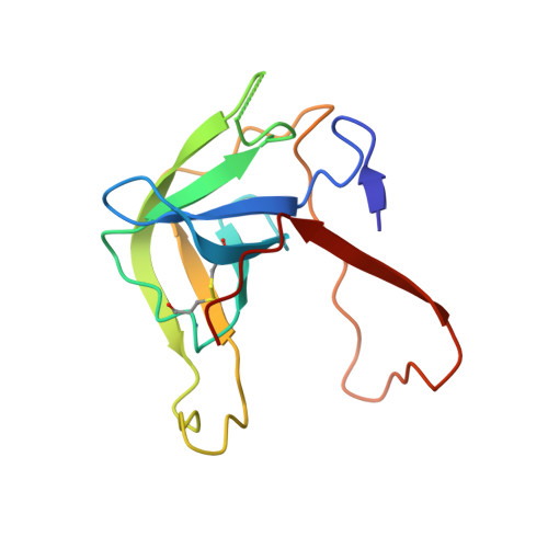
CHYMOTRYPSINOGEN B CHAIN B128Rattus norvegicusMutation(s): 0 
EC: 3.4.21.1
UniProt
Find proteins for P07338 (Rattus norvegicus)
Explore P07338 
Go to UniProtKB:  P07338
Entity Groups  
Sequence Clusters30% Identity50% Identity70% Identity90% Identity95% Identity100% Identity
UniProt GroupP07338
Sequence Annotations
Expand
  • Reference Sequence
Find similar proteins by:  (by identity cutoff)  |  3D Structure
Entity ID: 3
MoleculeChains Sequence LengthOrganismDetailsImage
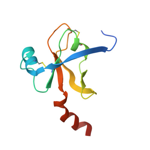
CHYMOTRYPSINOGEN B CHAIN C99Rattus norvegicusMutation(s): 2 
EC: 3.4.21.1
UniProt
Find proteins for P07338 (Rattus norvegicus)
Explore P07338 
Go to UniProtKB:  P07338
Entity Groups  
Sequence Clusters30% Identity50% Identity70% Identity90% Identity95% Identity100% Identity
UniProt GroupP07338
Sequence Annotations
Expand
  • Reference Sequence
Experimental Data & Validation

Experimental Data

  • Method: X-RAY DIFFRACTION
  • Resolution: 2.20 Å
  • R-Value Free: 0.334 
  • R-Value Work: 0.276 
  • R-Value Observed: 0.279 
  • Space Group: P 1 21 1
Unit Cell:
Length ( Å )Angle ( ˚ )
a = 34.69α = 90
b = 64.351β = 102.09
c = 44.224γ = 90
Software Package:
Software NamePurpose
REFMACrefinement
MOSFLMdata reduction
SCALAdata scaling
AMoREphasing

Structure Validation

View Full Validation Report


Entry History 

Deposition Data

Revision History  (Full details and data files)

  • Version 1.0: 2007-09-18
    Type: Initial release
  • Version 1.1: 2011-07-13
    Changes: Advisory, Version format compliance
  • Version 1.2: 2019-03-06
    Changes: Advisory, Data collection, Experimental preparation, Other
  • Version 1.3: 2019-05-08
    Changes: Data collection, Experimental preparation
  • Version 1.4: 2023-12-13
    Changes: Advisory, Data collection, Database references, Other, Refinement description