Structural and Mutagenic Analysis of Foot-and-Mouth Disease Virus 3C Protease Reveals the Role of the {Beta}-Ribbon in Proteolysis.
Sweeney, T.R., Roque-Rosell, N., Birtley, J.R., Leatherbarrow, R.J., Curry, S.(2007) J Virol 81: 115
- PubMed: 17065215
- DOI: https://doi.org/10.1128/JVI.01587-06
- Primary Citation of Related Structures:
2J92 - PubMed Abstract:
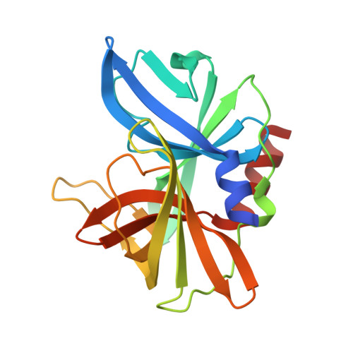
The 3C protease (3C(pro)) from foot-and-mouth disease virus (FMDV), the causative agent of a widespread and economically devastating disease of domestic livestock, is a potential target for antiviral drug design. We have determined the structure of a new crystal form of FMDV 3C(pro), a chymotrypsin-like cysteine protease, which reveals features that are important for catalytic activity. In particular, we show that a surface loop which was disordered in previous structures adopts a beta-ribbon structure that is conformationally similar to equivalent regions on other picornaviral 3C proteases and some serine proteases. This beta-ribbon folds over the peptide binding cleft and clearly contributes to substrate recognition. Replacement of Cys142 at the tip of the beta-ribbon with different amino acids has a significant impact on enzyme activity and shows that higher activity is obtained with more hydrophobic side chains. Comparison of the structure of FMDV 3C(pro) with homologous enzyme-peptide complexes suggests that this correlation arises because the side chain of Cys142 contacts the hydrophobic portions of the P2 and P4 residues in the peptide substrate. Collectively, these findings provide compelling evidence for the role of the beta-ribbon in catalytic activity and provide valuable insights for the design of FMDV 3C(pro) inhibitors.
Organizational Affiliation:
Biophysics Section, Blackett Laboratory, Imperial College, London SW7 2AZ, United Kingdom.