2J4W

Structure of a Plasmodium vivax apical membrane antigen 1-Fab F8.12.19 complex


Experimental Data Snapshot

  • Method: X-RAY DIFFRACTION
  • Resolution: 2.50 Å
  • R-Value Free: 0.241 
  • R-Value Work: 0.187 
  • R-Value Observed: 0.190 

wwPDB Validation   3D Report Full Report


This is version 2.2 of the entry. See complete history


Literature

Cross-Reactivity Studies of an Anti-Plasmodium Vivax Apical Membrane Antigen 1 Monoclonal Antibody: Binding and Structural Characterisation.

Igonet, S.Vulliez-Le Normand, B.Faure, G.Riottot, M.M.Kocken, C.H.M.Thomas, A.W.Bentley, G.A.

(2007) J Mol Biol 366: 1523

  • DOI: https://doi.org/10.1016/j.jmb.2006.12.028

  • PubMed Abstract: 

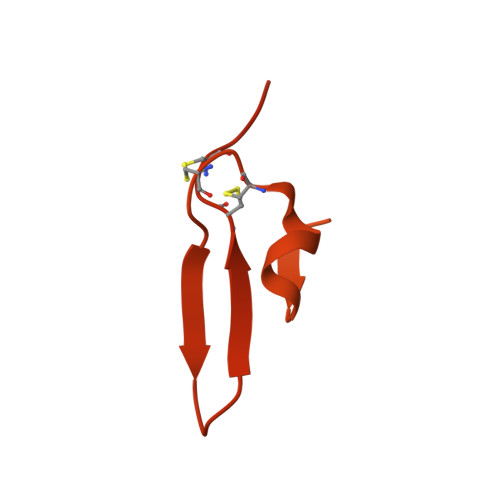
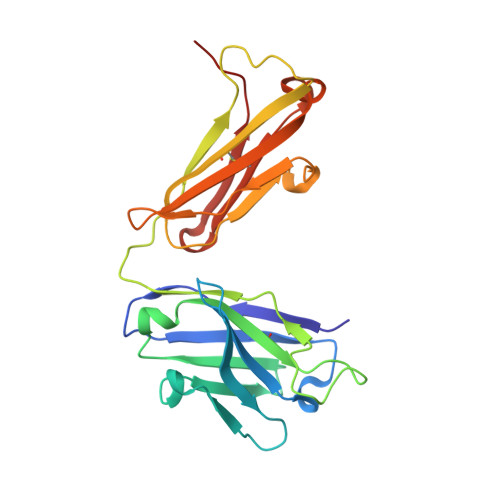
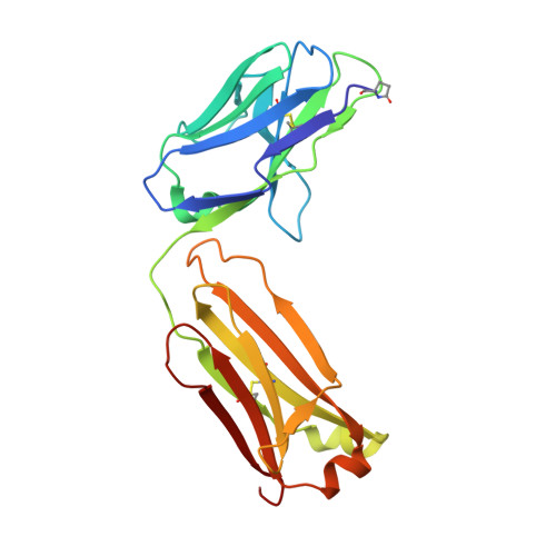
    Apical membrane antigen 1 (AMA1) has an important, but as yet uncharacterised, role in host cell invasion by the malaria parasite, Plasmodium. The protein, which is quite conserved between Plasmodium species, comprises an ectoplasmic region, a single transmembrane segment and a small cytoplasmic domain. The ectoplasmic region, which can induce protective immunity in animal models of human malaria, is a leading vaccine candidate that has entered clinical trials. The monoclonal antibody F8.12.19, raised against the recombinant ectoplasmic region of AMA1 from Plasmodium vivax, cross-reacts with homologues from Plasmodium knowlesi, Plasmodium cynomolgi, Plasmodium berghei and Plasmodium falciparum, as shown by immunofluorescence assays on mature schizonts. The binding of F8.12.19 to recombinant AMA1 from both P. vivax and P. falciparum was measured by surface plasmon resonance, revealing an apparent affinity constant that is about 100-fold weaker for the cross-reacting antigen when compared to the cognate antigen. Crystal structure analysis of Fab F8.12.19 complexed to AMA1 from P. vivax and P. falciparum shows that the monoclonal antibody recognises a discontinuous epitope located on domain III of the ectoplasmic region, the major component being a loop containing a cystine knot. The structures provide a basis for understanding the cross-reactivity. Antibody contacts are made mainly to main-chain and invariant side-chain atoms of AMA1; contact antigen residues that differ in sequence are located at the periphery of the antigen-binding site and can be accommodated at the interface between the two components of the complex. The implications for AMA1 vaccine development are discussed.


  • Organizational Affiliation

    Unité d'Immunologie Structurale, CNRS URA 2185, Département de Biologie Structurale et Chimie, Institut Pasteur, 25 rue du Dr. Roux, 75724 Paris cedex 15, France.


Macromolecules
Find similar proteins by:  (by identity cutoff)  |  3D Structure
Entity ID: 1
MoleculeChains Sequence LengthOrganismDetailsImage
APICAL MEMBRANE ANTIGEN 1A [auth D]445Plasmodium vivaxMutation(s): 3 
UniProt
Find proteins for Q9TY14 (Plasmodium vivax)
Explore Q9TY14 
Go to UniProtKB:  Q9TY14
Entity Groups  
Sequence Clusters30% Identity50% Identity70% Identity90% Identity95% Identity100% Identity
UniProt GroupQ9TY14
Sequence Annotations
Expand
  • Reference Sequence
Find similar proteins by:  (by identity cutoff)  |  3D Structure
Entity ID: 2
MoleculeChains Sequence LengthOrganismDetailsImage
FAB FRAGMENT OF MONOCLONAL ANTIBODY F8.12.19B [auth H]225Mus musculusMutation(s): 0 
UniProt
Find proteins for Q569B4 (Rattus norvegicus)
Explore Q569B4 
Go to UniProtKB:  Q569B4
Entity Groups  
Sequence Clusters30% Identity50% Identity70% Identity90% Identity95% Identity100% Identity
UniProt GroupQ569B4
Sequence Annotations
Expand
  • Reference Sequence
Find similar proteins by:  (by identity cutoff)  |  3D Structure
Entity ID: 3
MoleculeChains Sequence LengthOrganismDetailsImage
FAB FRAGMENT OF MONOCLONAL ANTIBODY F8.12.19C [auth L]213Mus musculusMutation(s): 0 
UniProt
Find proteins for Q5XFY8 (Mus musculus)
Explore Q5XFY8 
Go to UniProtKB:  Q5XFY8
Entity Groups  
Sequence Clusters30% Identity50% Identity70% Identity90% Identity95% Identity100% Identity
UniProt GroupQ5XFY8
Sequence Annotations
Expand
  • Reference Sequence
Small Molecules
Modified Residues  1 Unique
IDChains TypeFormula2D DiagramParent
PCA
Query on PCA
C [auth L]L-PEPTIDE LINKINGC5 H7 N O3GLN
Experimental Data & Validation

Experimental Data

  • Method: X-RAY DIFFRACTION
  • Resolution: 2.50 Å
  • R-Value Free: 0.241 
  • R-Value Work: 0.187 
  • R-Value Observed: 0.190 
  • Space Group: P 63
Unit Cell:
Length ( Å )Angle ( ˚ )
a = 171.787α = 90
b = 171.787β = 90
c = 44.748γ = 120
Software Package:
Software NamePurpose
REFMACrefinement
XDSdata reduction
XSCALEdata scaling
AMoREphasing

Structure Validation

View Full Validation Report


Entry History 

Deposition Data

Revision History  (Full details and data files)

  • Version 1.0: 2007-01-30
    Type: Initial release
  • Version 1.1: 2011-05-08
    Changes: Version format compliance
  • Version 1.2: 2011-07-13
    Changes: Version format compliance
  • Version 2.0: 2020-03-11
    Changes: Derived calculations, Other, Polymer sequence
  • Version 2.1: 2020-03-18
    Changes: Derived calculations
  • Version 2.2: 2023-12-13
    Changes: Data collection, Database references, Refinement description